Advanced Human Reproductive Physiology
Gamete Maturation Signaling
Oocyte Maturation: Breaking the Arrest
The journey of an oocyte is one of prolonged patience followed by a rapid burst of activity. Mammalian oocytes are held in meiotic arrest at prophase I, a state known as the dictyate stage, for potentially years. This cellular stasis is not passive; it's actively maintained by a sophisticated signaling network within the cumulus-oocyte complex (COC). The surrounding cumulus cells transport cyclic GMP (cGMP) into the oocyte through gap junctions. This influx of cGMP is critical.
Inside the oocyte, cGMP inhibits the enzyme phosphodiesterase 3A (PDE3A). With PDE3A suppressed, cyclic AMP (cAMP) levels remain high. This elevated cAMP activates Protein Kinase A (PKA), a key player in maintaining the arrest. Active PKA phosphorylates and inactivates components of the cell cycle machinery, like CDC25B and Wee1 kinase, which are necessary for activating the Maturation Promoting Factor (MPF). As long as MPF is dormant, the oocyte remains paused in prophase I.
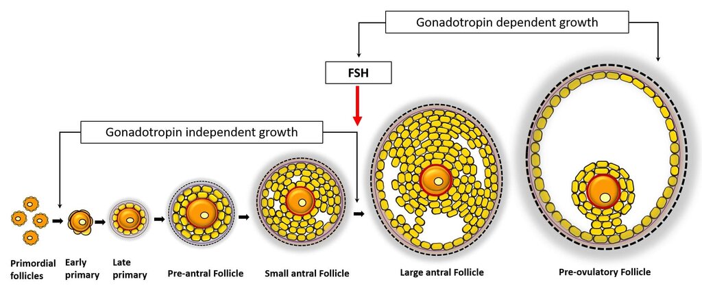
Meiotic resumption is triggered by the luteinizing hormone (LH) surge. This hormonal signal causes the gap junctions between cumulus cells and the oocyte to close. The supply of cGMP is cut off, and its concentration within the oocyte plummets. This frees PDE3A to begin its work: rapidly hydrolyzing cAMP. As cAMP levels fall, PKA activity decreases, lifting the brakes on the cell cycle machinery. The result is the swift activation of MPF, which initiates Germinal Vesicle Breakdown (GVB), the dissolution of the nuclear envelope. This marks the end of meiotic arrest and the transition into meiosis II. It's a precisely timed cascade that releases the oocyte from its long-held stasis.
Crucially, oocyte maturation isn't a single event. It involves two distinct, but ideally coordinated, processes: nuclear maturation and cytoplasmic maturation. Nuclear maturation is the visible progression of chromosomes through meiosis, ending with the extrusion of the first polar body and arrest at metaphase II. Cytoplasmic maturation is a less obvious but equally vital process where the oocyte's cytoplasm reorganizes. This involves redistributing organelles, synthesizing proteins, and stockpiling mRNAs and other molecules that will guide the earliest stages of embryonic development post-fertilization. These two processes can become uncoupled, leading to an oocyte that is chromosomally mature but cytoplasmically incompetent, often resulting in failed fertilization or developmental arrest.
Sperm Maturation: A Tale of Remodeling and Activation
The maturation of sperm is a dramatic transformation known as spermiogenesis. This process fundamentally remodels the spermatid, turning a round, conventional-looking cell into a streamlined, motile spermatozoon. A key aspect of this is extreme chromatin condensation, which is necessary to protect the paternal genome and create a hydrodynamically efficient head.
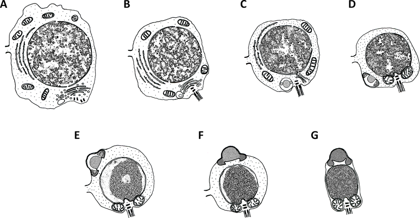
This condensation is a two-step process. First, histones, the usual protein spools for DNA, are replaced by smaller, more basic transition proteins. Shortly after, these are themselves replaced by even smaller, highly basic proteins called protamines. Protamines, rich in arginine and cysteine, bind tightly to the DNA backbone, neutralizing its charge and allowing it to be packaged into an incredibly dense, almost crystalline state. This hyper-condensation silences nearly all transcription and renders the DNA exceptionally stable.
But spermiogenesis is just the beginning. Sperm released from the testes are not yet capable of fertilization. They must undergo further maturation in the epididymis, a long, coiled tube. Here, they acquire motility and undergo significant changes to their plasma membrane, including the addition and modification of various proteins and lipids. This process primes them for the final step: capacitation.
Capacitation is the final gauntlet. It's a series of physiological changes sperm undergo in the female reproductive tract that makes them competent to fertilize an egg.
Two key molecular events define capacitation. The first is cholesterol efflux. The sperm membrane is initially rich in cholesterol, which makes it rigid. As sperm travel through the female tract, albumin and other molecules strip this cholesterol away. This increases membrane fluidity, which is essential for the later acrosome reaction. The second event is protein phosphorylation, which triggers hyperactivation. This is a shift in motility from a steady, forward progression to a frantic, whip-like motion. Hyperactivated motility gives the sperm the power needed to detach from the oviduct wall and penetrate the egg's protective layers. Together, these carefully orchestrated signaling events ensure that only the most robust and ready sperm reach their destination.