Advanced Human Circulation and Fluid Dynamics
Cardiac Dynamics
The Heart's Inner Spark
Unlike skeletal muscles that wait for a signal from the brain, the heart generates its own electrical rhythm. This property is called , meaning the heartbeat originates within the heart muscle itself. It doesn't need external nerves to initiate a beat, though the nervous system can certainly influence its rate, telling it to speed up or slow down.
The process begins in the sinoatrial (SA) node, a small cluster of cells in the right atrium. These are the pacemaker cells. They spontaneously generate electrical impulses, called action potentials, at a regular pace. This initial spark sets the tempo for the entire heart.
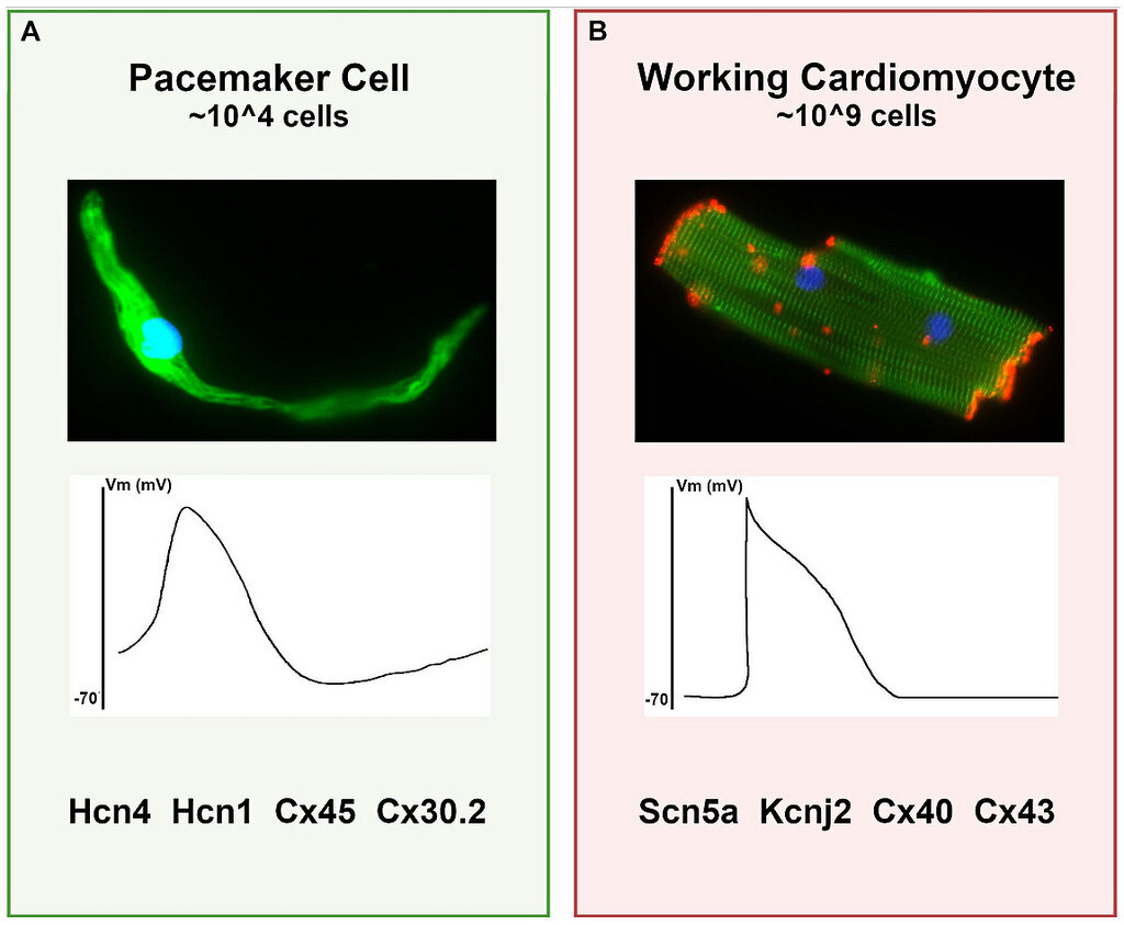
An action potential in a cardiac muscle cell is a rapid change in voltage across the cell membrane. It occurs in distinct phases driven by the flow of ions like sodium (), potassium (), and calcium () through specific channels. First, sodium ions rush into the cell, causing a rapid depolarization (the upstroke). This is followed by a plateau phase, unique to cardiac muscle, where calcium ions enter the cell. This influx of calcium is the crucial link that triggers the mechanical contraction of the muscle fibres. Finally, potassium ions exit the cell, causing it to repolarize and return to its resting state, ready for the next beat.
The Conduction Highway
For the heart to pump efficiently, this electrical signal must spread rapidly and in a highly coordinated manner. It can't just be a chaotic wave. The heart has a specialized conduction system, a network of pathways that acts like electrical wiring.
The conduction system within the heart is vital because it permits a rapid and organized depolarization of ventricular myocytes that is necessary for the efficient generation of pressure during systole.
From the SA node, the impulse spreads across both atria, causing them to contract and push blood into the ventricles. The signal then reaches the atrioventricular (AV) node, located between the atria and ventricles. The AV node acts as a gatekeeper, briefly delaying the signal. This pause is critical; it gives the ventricles time to fill completely with blood before they are told to contract.
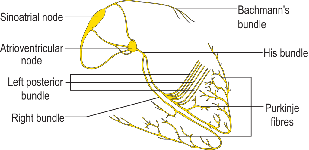
After the delay, the signal travels down the Bundle of His, which splits into right and left bundle branches. These branches extend through the septum towards the apex of the heart. The signal finally spreads throughout the ventricular walls via a network of , causing the ventricles to contract from the bottom up. This powerful, coordinated squeeze ejects blood into the pulmonary artery and the aorta.
Pressure and Volume
The entire sequence of electrical and mechanical events is known as the cardiac cycle. It consists of two main phases: diastole (relaxation and filling) and systole (contraction and ejection). The relationship between pressure and volume within a ventricle during one complete cycle can be visualised with a pressure-volume loop.
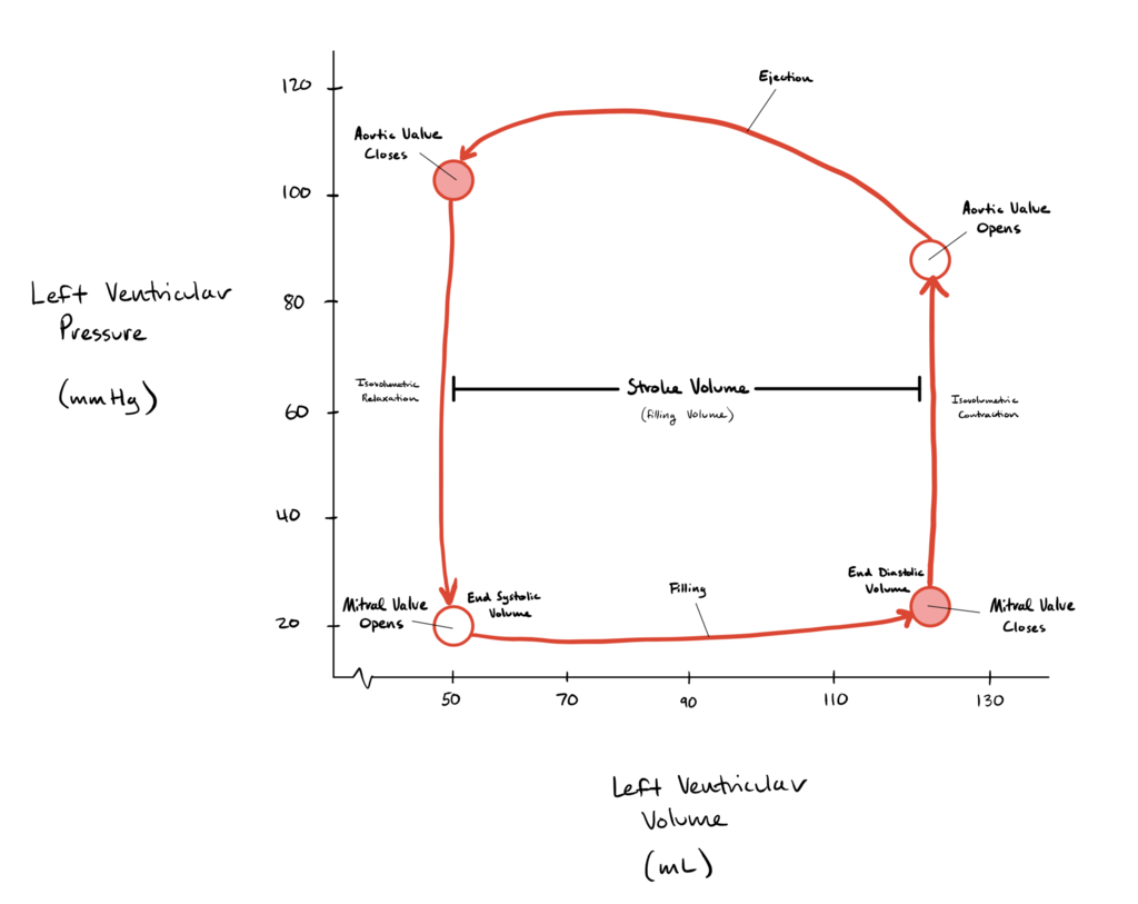
This loop provides a powerful summary of cardiac function. Let's break down the four distinct phases of the cycle as seen in the left ventricle:
| Phase | Description |
|---|---|
| 1. Ventricular Filling | The mitral valve is open, and the ventricle relaxes and fills with blood from the atrium. Volume increases significantly, but pressure rises only slightly. |
| 2. Isovolumetric Contraction | Both the mitral and aortic valves are closed. The ventricle contracts, causing a sharp increase in pressure, but the volume of blood inside does not change. |
| 3. Ventricular Ejection | Pressure in the ventricle exceeds pressure in the aorta, forcing the aortic valve to open. Blood is rapidly ejected, causing ventricular volume to decrease. |
| 4. Isovolumetric Relaxation | The aortic valve closes. The ventricle relaxes, and pressure falls dramatically. Both valves are closed again, so volume remains constant at its lowest point. The cycle restarts when ventricular pressure drops below atrial pressure, causing the mitral valve to open. |
This elegant integration of electrical signals, coordinated muscle contraction, and valve mechanics ensures that with every beat, the heart effectively pumps blood to the lungs and the rest of the body.
Time to check your understanding of how the heart's electrical and mechanical events are linked.
What is the term for the heart's intrinsic ability to generate its own electrical rhythm without needing signals from the nervous system?
What is the primary function of the atrioventricular (AV) node in the heart's conduction system?