Advanced Human Anatomy and Systemic Integration
Thoracic Cavity Integration
The Pressurized Chamber
The thoracic cavity is more than just a bony cage. It's a dynamic, pressurized system where the heart, lungs, and diaphragm work in tight coordination. The key to this entire operation is the pleural space—the thin, fluid-filled gap between the lungs and the chest wall. This space maintains a negative pressure relative to the atmosphere, acting like a gentle suction that keeps the lungs tethered to the inside of the rib cage. When the chest wall expands, the lungs are pulled along with it, forcing them to inflate.
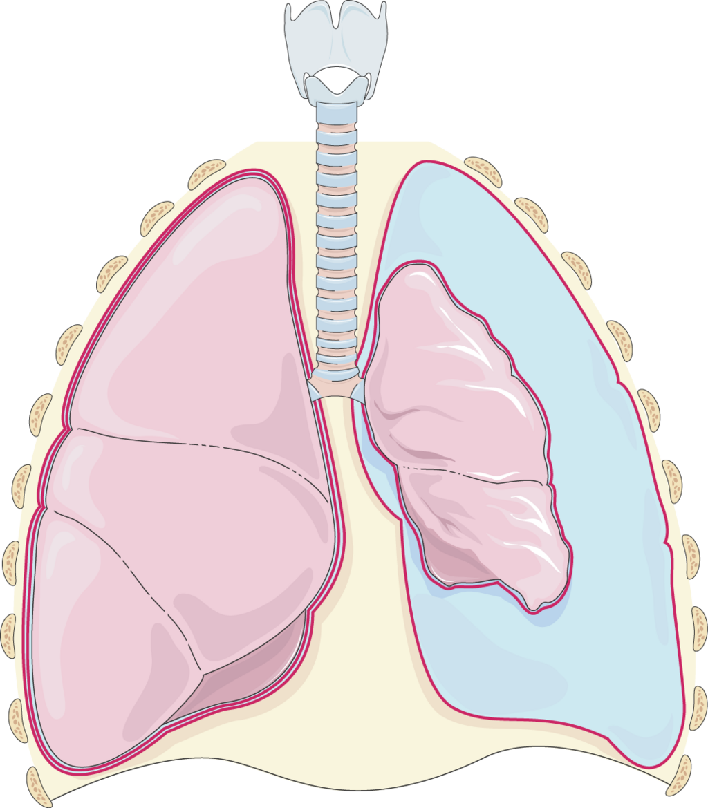
This delicate pressure balance is critical. If air breaches the pleural space, this suction is lost, and the lung’s natural elasticity causes it to collapse—a condition called a pneumothorax. In a more dangerous scenario, a occurs when a flap of tissue creates a one-way valve. Air enters the pleural space with each breath but can't escape, causing pressure to build rapidly. This doesn't just collapse the lung; it shoves the entire central compartment of the chest, the mediastinum, to the opposite side, compressing the heart and major blood vessels. This severely obstructs blood from returning to the heart, leading to a rapid drop in blood pressure and circulatory collapse.
The Rhythmic Squeeze
The diaphragm, a large dome-shaped muscle at the base of the thoracic cavity, is the primary engine of breathing. When it contracts, it flattens and moves downward, dramatically increasing the vertical dimension of the chest. This expansion drops the pressure inside the cavity, pulling air into the lungs. This isn't just a respiratory event; it's a circulatory one, too.
As diaphragm is pulled down, volume of lungs increases. Air pressure in lungs drop. So air rushes from the outside, down into lungs, until the pressure inside = pressure outside
The drop in intrathoracic pressure also lowers the pressure within the heart's right atrium. This pressure gradient between the abdomen and the chest actively sucks deoxygenated blood from the lower body up into the heart. This
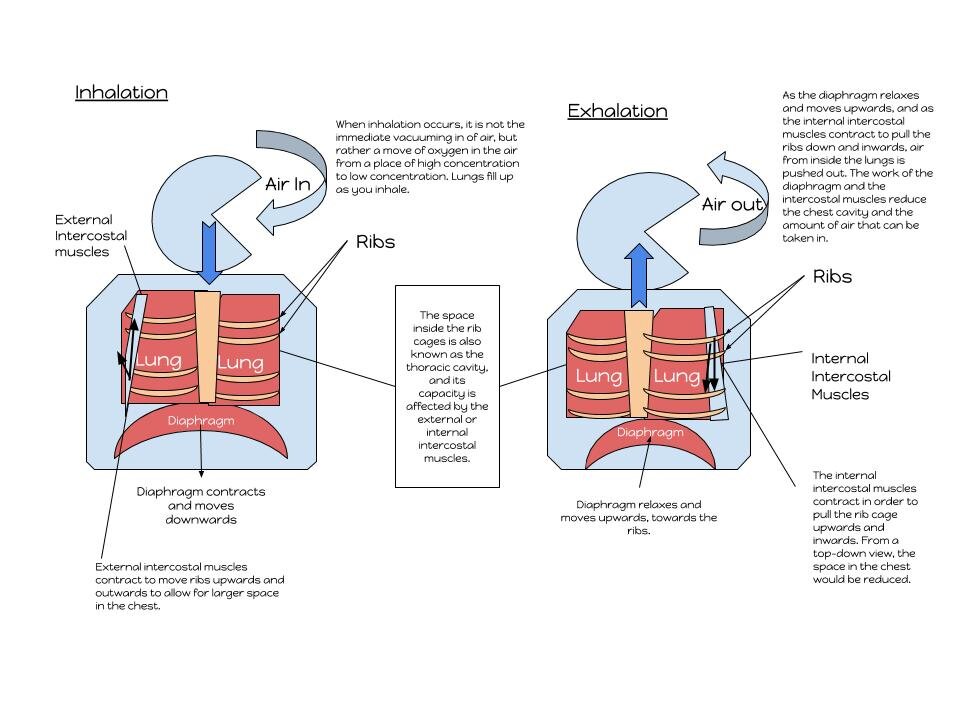
This “thoracic pump” mechanism is a prime example of cardiopulmonary integration, where the simple act of breathing directly assists in cardiac filling. The reverse happens during exhalation. As the diaphragm relaxes and rises, intrathoracic pressure increases, which helps the left ventricle pump oxygenated blood out to the body.
A Matter of Space
The heart, lungs, and major vessels are packed together within the and are subject to tight spatial constraints. The heart's ability to fill and pump is directly affected by the space available. In healthy states, this isn't an issue. But in conditions like chronic obstructive pulmonary disease (COPD), the lungs become hyperinflated and don't fully deflate. They essentially take up more space than they should.
This chronic over-inflation physically compresses the heart and increases the pressure in the pulmonary arteries. The right ventricle must work much harder to pump blood into these high-pressure lungs, a condition known as right-sided heart strain, or cor pulmonale. This demonstrates how a primary lung disease can directly cause heart failure, not through a circulatory problem, but through a biomechanical and spatial one.
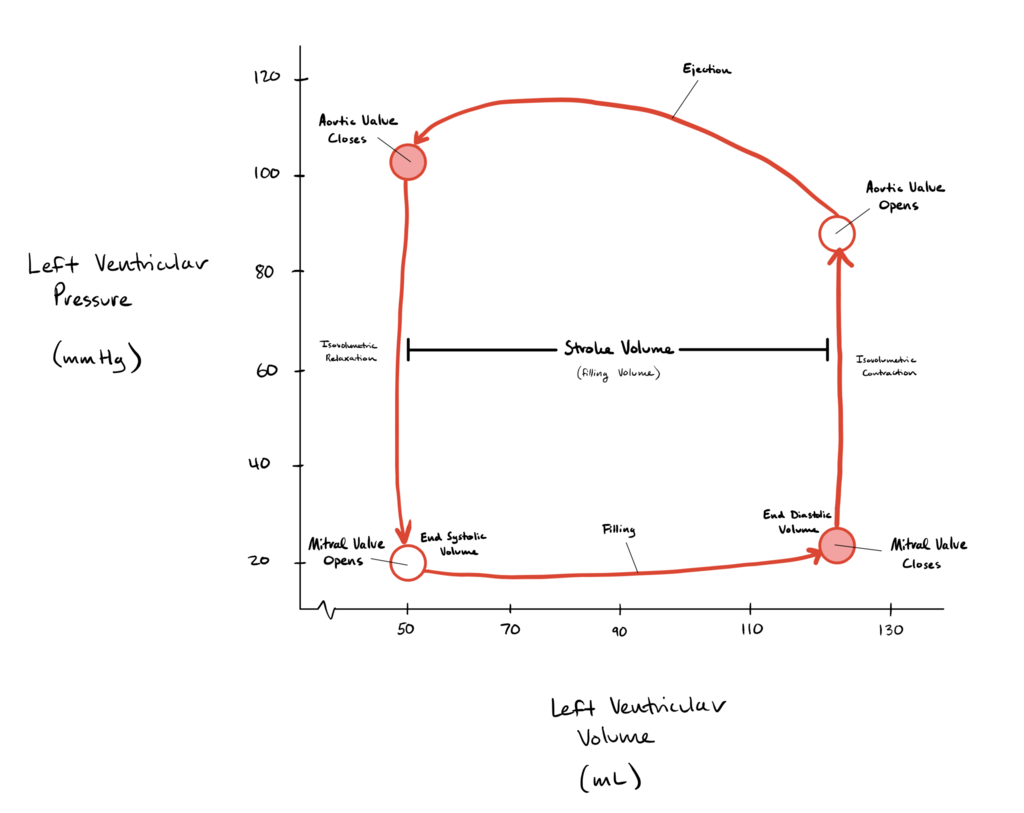
Similarly, the compliance of the thoracic wall—its ability to stretch and expand—plays a role. In conditions where the chest wall is stiff, such as kyphoscoliosis or in obese patients, the work of breathing increases. This reduces the efficiency of the thoracic pump and can limit the heart's ability to fill properly, affecting overall cardiac output.
Time to check your understanding of how this integrated system works.
What is the primary function of the negative pressure within the pleural space?
What is the key feature that distinguishes a tension pneumothorax from a simple pneumothorax?
Understanding these mechanical relationships is key to seeing the thoracic cavity not as a collection of parts, but as a single, elegant machine.