Advanced Functions of the Human Spleen
Internal Splenic Architecture
A Filter in Two Colours
The spleen's power lies in its sophisticated internal design. Far from being a simple organ, it's organised into two distinct, interconnected regions: the white pulp and the red pulp. Each has a specific job, and together they make the spleen a master of blood filtration and immune surveillance.
Imagine slicing the spleen open. You'd see a dark red, spongy mass (the red pulp) dotted with small, greyish-white islands of tissue (the white pulp). This isn't random. The entire structure is supported by a network of fibrous partitions called trabeculae, which extend inwards from the outer capsule. These trabeculae act like a scaffold, carrying the main blood vessels deep into the organ.
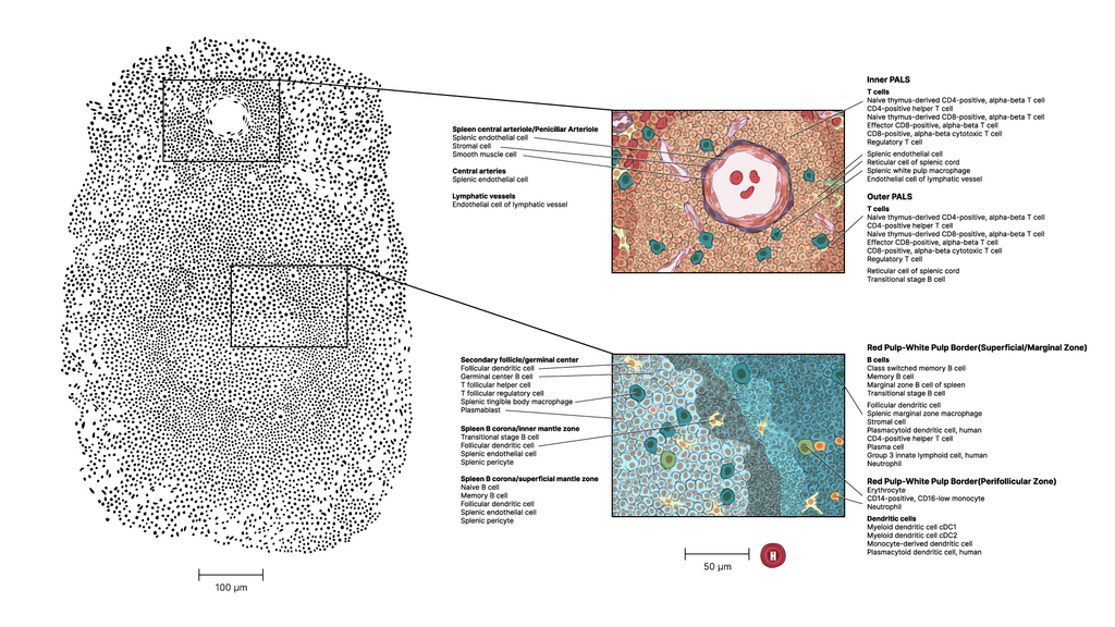
White Pulp: The Immune Hub
The white pulp is the spleen's lymphatic component, acting as a crucial meeting point for immune cells. It's strategically built around the arteries entering the spleen. As arterial blood flows in, it first passes through these immune-rich zones. The central feature of the white pulp is the (PALS), which is a dense sleeve of T-cells that wraps around the central arteries.
Embedded within or attached to the PALS are lymphoid follicles, which are rich in B-cells. This is where B-cells are activated and mature, ready to produce antibodies against invaders detected in the blood. The close proximity of T-cells in the PALS and B-cells in the follicles allows them to communicate effectively and mount a coordinated immune response.
Red Pulp and the Marginal Zone
Surrounding the white pulp is the red pulp, which makes up most of the spleen's tissue. This is the filtration plant. The red pulp consists of two key parts: splenic cords and venous sinuses. The splenic cords, also known as the , are a meshwork of reticular fibres packed with immune cells, especially macrophages.
The venous sinuses are like wide, leaky blood vessels. Blood that successfully navigates the splenic cords must pass through the walls of these sinuses to re-enter circulation. This process provides a final check, removing cellular debris and pathogens from the blood.
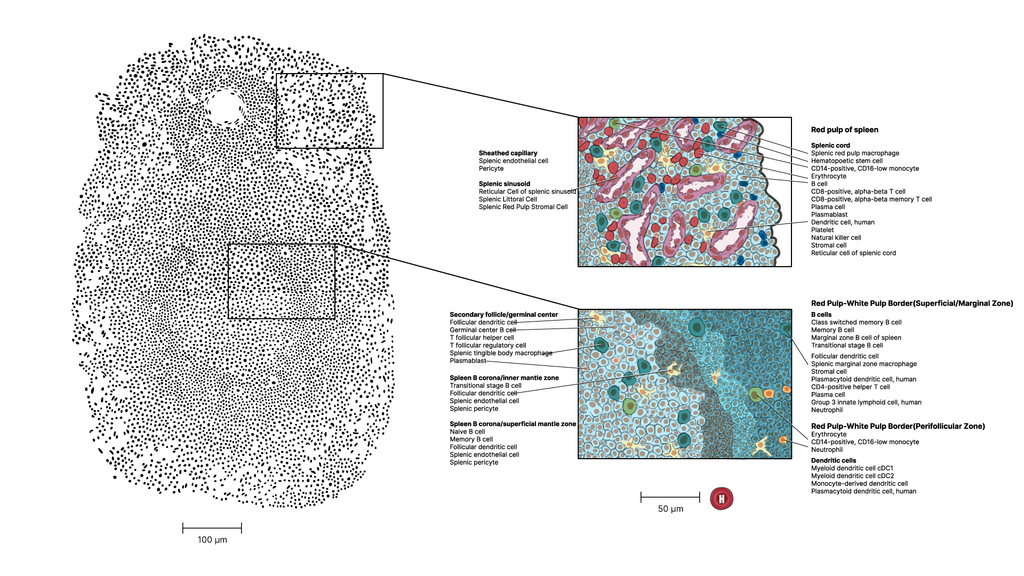
A crucial area is the marginal zone, which sits at the interface between the red and white pulp. This is a strategic location that receives a large amount of blood flow. It acts as a primary site for capturing blood-borne antigens and presenting them to the immune cells in the white pulp. The marginal zone is rich in specialized macrophages and B-cells, making it a first line of defence against pathogens entering the bloodstream.
In essence, the white pulp is for immune recognition, while the red pulp handles the physical cleanup of the blood. The marginal zone acts as the bridge connecting the two functions.
And with that, let's test what you've learned about the spleen's internal world.
What are the two primary, distinct regions that make up the internal structure of the spleen?
The Periarteriolar Lymphoid Sheath (PALS), a key component of the white pulp, is primarily composed of which type of immune cell?
The spleen's intricate architecture allows it to serve as a dynamic link between the circulatory and immune systems, constantly monitoring and maintaining the health of our blood.