Advanced Functions of the Human Liver
Hepatic Microanatomy and Bloodflow
The Functional Units of the Liver
While the liver appears as a single, large organ from the outside, its internal structure is a marvel of microscopic organization. The classical way to describe this structure is through the liver lobule, a hexagonal unit of tissue. Imagine millions of these tiny hexagons packed together, each organized around a central vein, like tiles on a floor.
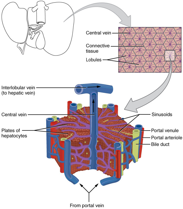
At each corner of the hexagon lies a portal triad, a trio of vessels consisting of a branch of the hepatic artery, a branch of the hepatic portal vein, and a bile duct. This arrangement is key to the liver's function. Blood flows from the triads at the periphery, moves inward through specialized capillaries called sinusoids, and drains into the central vein.
However, a more functionally relevant model is the liver acinus. Instead of a hexagon, the acinus is a diamond-shaped area of tissue centered on the blood supply line running between two portal triads. This model helps explain how liver cells, or hepatocytes, function based on their proximity to incoming blood.
Metabolic Zoning
The liver has a unique dual blood supply. The hepatic portal vein delivers nutrient-rich, but oxygen-poor, blood from the digestive system. The hepatic artery delivers oxygen-rich blood from the heart. These two sources mix within the liver's sinusoids, creating a gradient of oxygen and nutrients across the acinus.
This gradient divides the acinus into three metabolic zones:
| Zone | Location | Oxygen/Nutrient Level | Primary Functions |
|---|---|---|---|
| Zone 1 | Closest to the portal triad | Highest | Oxidative metabolism, urea synthesis, gluconeogenesis |
| Zone 2 | Intermediate zone | Medium | Transitional functions |
| Zone 3 | Closest to the central vein | Lowest | Glycolysis, lipogenesis, drug detoxification (cytochrome P450) |
This zoning has important clinical implications. Zone 1 is the first to receive blood, so it's most vulnerable to certain viral toxins that don't require activation. In contrast, Zone 3 cells are the most susceptible to damage from hypoxia (low oxygen) and to toxins that are activated by the P450 enzyme system, such as the active metabolite of acetaminophen.
The Exchange Floor
Blood doesn't just wash over the hepatocytes. It flows through the sinusoids, which are capillaries with a unique, fenestrated (windowed) endothelium. These large pores allow blood plasma, but not red blood cells, to filter out and come into direct contact with the hepatocytes.
This exchange happens in a narrow region called the , which lies between the sinusoidal endothelium and the hepatocytes. This is where the liver does its real work: absorbing nutrients, releasing glucose, and processing toxins.
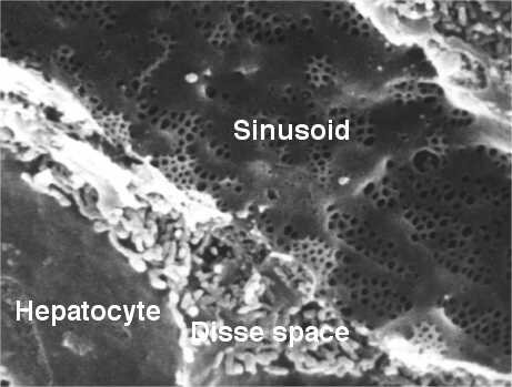
Within the sinusoids and the Space of Disse reside other crucial cells. Kupffer cells are resident macrophages that act as the liver's first line of immune defense, engulfing pathogens and cellular debris from the portal blood. Hepatic stellate cells are also found here, playing a key role in vitamin A storage and, in cases of injury, scar tissue formation.
Think of the Space of Disse as the loading dock of the liver, where raw materials from the blood are offloaded for processing by the hepatocyte factory workers.
Bile's Journey Out
While blood flows inward toward the central vein, the flow of bile moves in the opposite direction. Hepatocytes produce bile and secrete it into tiny channels called bile canaliculi that run between them. These canaliculi are like microscopic gutters.
They collect the bile and drain it outward, toward the portal triads. There, they merge into the larger bile ducts. This intricate network, known as the biliary tree, eventually carries bile out of the liver for storage in the gallbladder or for immediate use in digestion.
Let's review the key terms we've covered.
Now, test your understanding of these concepts.
What are the three components of a hepatic portal triad?
In the liver acinus model, which zone is most susceptible to damage from hypoxia (low oxygen)?
Understanding this microscopic architecture is fundamental to grasping how the liver performs its hundreds of vital functions and how different diseases can selectively damage this resilient organ.