Advanced Clinical Cardiology Management
Advanced Multimodality Imaging
Beyond the Echocardiogram
As cardiologists, we rely on echocardiography for its immediate, invaluable insights. Yet, many clinical puzzles remain unsolved by standard imaging. When a patient presents with chest pain but clean coronary arteries, or when heart failure has an elusive cause, we need to look deeper. This is where advanced multimodality imaging, particularly Cardiac Magnetic Resonance (CMR) and Positron Emission Tomography (PET), becomes essential.
These modalities move us from observing anatomy and function to characterizing tissue and quantifying physiology. The 2025 ACC Advanced Cardiovascular Imaging training statements underscore this shift, emphasizing the need for cardiologists to proficiently integrate CMR and hybrid PET/CT or PET/CMR to solve complex diagnostic challenges and guide targeted therapies.
CMR for Tissue Characterization
CMR's strength lies in its ability to non-invasively characterize myocardial tissue. Using techniques like T1 and T2 mapping for edema and fibrosis, and especially Late Gadolinium Enhancement (LGE), we can differentiate between pathologies that appear similar on other modalities. This is particularly powerful in the workup of (Myocardial Infarction with Non-Obstructive Coronary Arteries).
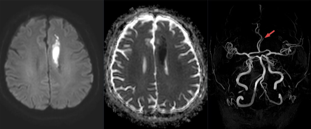
In a MINOCA patient, the pattern of LGE on CMR can pinpoint the cause. Subendocardial LGE points to a true ischemic event, like a ruptured plaque with spontaneous reperfusion. In contrast, mid-myocardial or epicardial LGE suggests myocarditis. The absence of LGE, coupled with wall motion abnormalities and edema, might indicate Takotsubo cardiomyopathy. Each pattern leads to a vastly different treatment path.
LGE patterns are the fingerprints of myocardial injury. A subendocardial pattern follows a vascular territory, while a non-coronary distribution suggests an infiltrative or inflammatory process.
This tissue-level insight extends to inflammatory and infiltrative cardiomyopathies. For suspected cardiac sarcoidosis, CMR is invaluable. The characteristic patchy, mid-myocardial, or epicardial LGE in the basal septum and lateral wall is highly suggestive. Similarly, in cardiac amyloidosis, a diffuse, subendocardial LGE pattern and markedly elevated native T1 values can strongly support the diagnosis, often prompting further specific testing.
PET for Function and Viability
While CMR excels at characterizing tissue structure, PET excels at quantifying metabolic function and absolute blood flow. Hybrid imaging with PET/CT or PET/CMR combines anatomical context with physiological data, providing a comprehensive assessment of coronary artery disease, especially in complex cases.
A key application is the assessment of (CMD), a common cause of ischemia in patients with non-obstructive coronary arteries. These patients often have normal stress tests using relative perfusion agents but continue to have symptoms. PET allows for the non-invasive quantification of Myocardial Blood Flow (MBF) and Myocardial Flow Reserve (MFR).
This quantitative approach moves beyond simply identifying perfusion defects. It provides a specific diagnosis of CMD, which carries prognostic weight and guides therapy toward agents that target the microvasculature.
PET is also the gold standard for assessing myocardial viability in patients with severe left ventricular dysfunction being considered for revascularization. By simultaneously imaging perfusion (with agents like Rubidium-82 or N-13 ammonia) and metabolism (with F-18 FDG), PET can identify hibernating myocardium. A region with reduced perfusion but preserved glucose metabolism (a perfusion-metabolism mismatch) indicates viable tissue that is likely to recover function after revascularization.
Integrating Modalities for Complex Cases
The true power of advanced imaging lies in selecting the right test for the right clinical question, often involving a combination of modalities.
For instance, assessing a prosthetic heart valve can be challenging with echocardiography alone due to acoustic shadowing. While CT provides excellent anatomical detail of the valve structure and can identify pannus or thrombus, it doesn't assess the hemodynamic impact. Here, a multimodality approach is key. Cine-fluoroscopy can assess leaflet mobility, and PET/CT can be used to detect infection by identifying areas of increased metabolic activity around the valve.
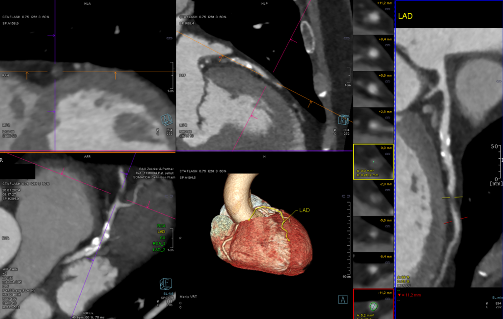
Another area of growing importance is 3D modeling for planning structural heart interventions. Data from high-resolution CT and 3D echocardiography are used to create precise, patient-specific models of cardiac anatomy. These models allow for virtual device implantation, helping to predict the fit of a transcatheter valve, plan access routes, and anticipate potential complications like coronary obstruction or paravalvular leak. Advanced strain imaging, particularly with speckle-tracking echocardiography, complements this by providing detailed analysis of regional myocardial function, which can influence procedural strategy and predict outcomes.
Test your understanding of these advanced imaging applications.
A patient with suspected Myocardial Infarction with Non-Obstructive Coronary Arteries (MINOCA) undergoes a Cardiac MRI (CMR). Which Late Gadolinium Enhancement (LGE) pattern is most indicative of an ischemic etiology, such as a plaque rupture with spontaneous reperfusion?
What is the primary advantage of using Positron Emission Tomography (PET) for evaluating coronary microvascular dysfunction (CMD) in patients with angina and non-obstructive coronary arteries?
By mastering these advanced modalities, we can move beyond one-size-fits-all diagnostics and provide more precise, effective care for our patients with complex cardiovascular disease.