Advanced Central Nervous System Architecture
Internal Brain Structures
The Deep Gray Matter
Beneath the folded cerebral cortex lies a collection of deep gray matter structures critical for motor control, sensory integration, and endocrine function. We will focus on two main groups: the basal nuclei and the diencephalon. These structures work in concert with the cortex to refine movement, process sensory information, and maintain the body's internal balance.
The Basal Nuclei
The basal nuclei, often called the basal ganglia, are a group of subcortical nuclei primarily involved in motor control. They don't initiate movement themselves, but rather modulate motor output from the cerebral cortex, ensuring movements are smooth, purposeful, and appropriately scaled. Think of them as a quality control system for your actions.
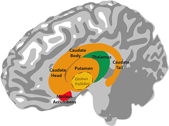
Anatomically, the major components are the caudate nucleus, putamen, and globus pallidus. The caudate nucleus has a distinctive C-shape that follows the curve of the lateral ventricle. It has a large head anteriorly, which tapers into a body and a slender tail.
The putamen and globus pallidus are located just lateral to the thalamus and are collectively shaped like a lens, earning them the name lentiform nucleus. The putamen is the more lateral part, while the globus pallidus is medial. The globus pallidus is further divided into an external segment (GPe) and an internal segment (GPi).
lentiform nucleus
noun
A lens-shaped mass of gray matter comprising the putamen and the globus pallidus, located deep within the cerebral hemispheres.
Functionally, these nuclei are often grouped differently. The —composed of the caudate nucleus and the putamen—serves as the main input station for the basal nuclei, receiving signals primarily from the cerebral cortex. The globus pallidus internus (GPi), along with the substantia nigra pars reticulata, acts as the primary output nucleus, sending inhibitory signals to the thalamus.
The interplay between these structures is crucial. Dysfunction in the basal nuclei pathways is central to movement disorders like Parkinson's disease and Huntington's disease.
The Internal Capsule
Weaving between these deep nuclei is the , a massive, V-shaped bundle of white matter fibres. It contains both ascending fibres carrying sensory information from the thalamus to the cortex (thalamocortical tracts) and descending fibres from the cortex to the brainstem and spinal cord (corticospinal and corticobulbar tracts). Its compact nature means that even a small lesion here can cause widespread motor and sensory deficits.
The internal capsule is divided into three main parts based on its relationship to the surrounding nuclei:
- Anterior limb: Situated between the head of the caudate nucleus and the lentiform nucleus.
- Genu: The 'knee' or bend of the capsule, located between the anterior and posterior limbs.
- Posterior limb: Found between the thalamus and the lentiform nucleus. This is the largest and most clinically significant part, as it contains the main motor and sensory fibres.
The Diencephalon
The diencephalon is a central brain region nestled between the cerebral hemispheres and the midbrain. It acts as a primary processing and relay centre for sensory information and autonomic control. Its major components are the thalamus, hypothalamus, and epithalamus.
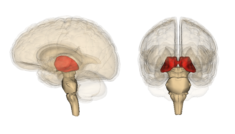
The thalamus is the largest part of the diencephalon, consisting of two egg-shaped masses of gray matter. It functions as the brain's main relay station. Almost all sensory information (except for olfaction) synapses in a specific thalamic nucleus before being relayed to the cerebral cortex. But it's more than a simple switchboard; it also processes and filters this information, playing a role in regulating consciousness, sleep, and alertness.
The hypothalamus lies inferior and anterior to the thalamus. Despite its small size, it is a vital control centre for the autonomic nervous system and the endocrine system, primarily through its connection to the pituitary gland. It regulates fundamental survival functions like body temperature, hunger, thirst, and circadian rhythms.
The epithalamus is located superior and posterior to the thalamus. Its main structures are the pineal gland, which secretes melatonin to regulate sleep-wake cycles, and the habenular nuclei, involved in emotional and motivational responses.
These structures form the walls of the third ventricle, a key landmark for identifying the diencephalon on neuroimaging. Understanding the precise anatomical and functional relationships between the basal nuclei, internal capsule, and diencephalon is fundamental to diagnosing and localising lesions in clinical neurology.
The lentiform nucleus is a collective term for which two structures?
A small lesion in the internal capsule is unlikely to cause significant motor or sensory deficits due to its small size.