Advanced Cardiac Mechanics and Physiology
Electrophysiology and Impulse Regulation
The Heart's Electrical Spark
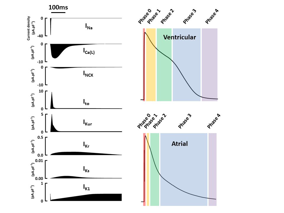
The heart's beat isn't just a simple muscle flex. It's a precisely timed electrical event, a wave of depolarization that sweeps through specialized cardiac cells. Unlike skeletal muscles that wait for a signal from the brain, the heart generates its own rhythm. This process, known as automaticity, is driven by the movement of ions across cell membranes.
Three key players are at work: sodium (), potassium (), and calcium (). At rest, cardiac cells have a negative charge inside relative to the outside. An electrical impulse begins when special channels open, allowing positively charged ions to rush in. This rapid influx of and then flips the cell's internal charge from negative to positive, a process called depolarization. This electrical change triggers the mechanical contraction of the muscle fiber. Shortly after, ions flow out, returning the cell to its negative resting state, or repolarization, allowing it to relax and prepare for the next beat.
The Chain of Command
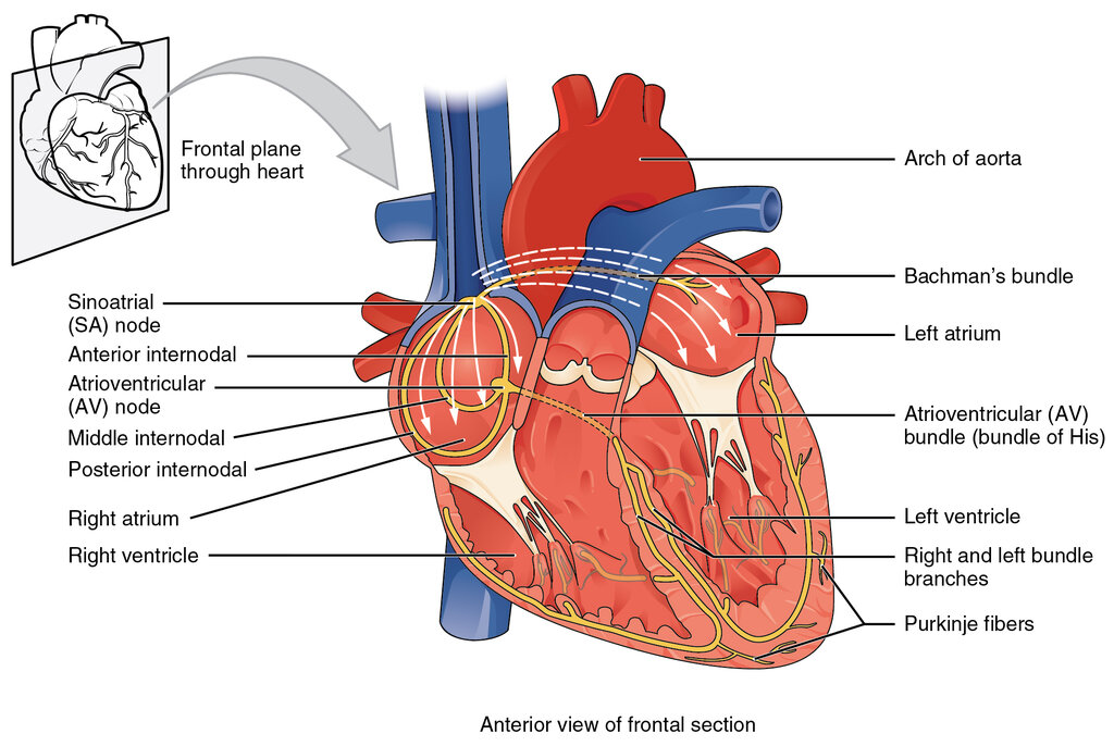
The heart's electrical system has a clear hierarchy. The main pacemaker is the Sinoatrial (SA) node, a small cluster of cells in the right atrium. It spontaneously depolarizes faster than any other part of the heart, typically firing 60-100 times per minute, setting the pace for the entire organ.
The signal spreads from the SA node across both atria, causing them to contract. It then travels to the Atrioventricular (AV) node, located near the center of the heart. If the SA node fails, the AV node can take over, but at a slower rate (40-60 beats per minute). Further down the line, other cells in the ventricles can act as a last resort, but their intrinsic rate is much slower, around 20-40 beats per minute. This principle, where the fastest pacemaker sets the rate and suppresses the slower ones, is called It's a built-in fail-safe system.
The Crucial Pause
When the electrical impulse reaches the AV node, something important happens: it slows down. This isn't a mistake; it's a critical design feature known as the AV delay. This pause, lasting about a tenth of a second, gives the atria enough time to finish contracting and push their full load of blood into the ventricles.
Without the AV delay, the atria and ventricles would contract at nearly the same time, severely compromising the heart's ability to pump blood effectively.
This delay is a result of the unique properties of the AV node's cells. They have fewer gap junctions, which are the channels that connect adjacent cells and allow electrical signals to pass. Fewer channels mean slower conduction. The action potentials in this region are also more dependent on the slower influx of calcium ions rather than the rapid rush of sodium ions seen elsewhere.
The Ventricular Superhighway
After the pause, the signal is ready to trigger the powerful ventricular contraction. The impulse travels from the AV node down the Bundle of His, which splits into right and left bundle branches. These branches then feed into a network of specialized fibers called Purkinje fibers that spread throughout the ventricular walls.
These fibers are the superhighway of the heart's electrical system. They conduct the electrical impulse up to four times faster than any other cardiac tissue. This incredible speed is crucial for coordinating the contraction of the ventricles. It ensures that all parts of the ventricular muscle depolarize almost simultaneously, leading to a powerful, unified squeeze from the bottom up. This wringing motion efficiently ejects blood into the aorta and pulmonary artery.
Reading the Signals
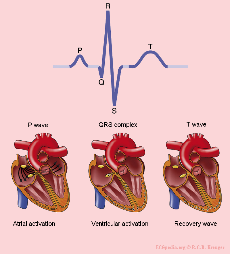
The sum of all this electrical activity can be measured on the skin's surface with an electrocardiogram, or ECG. Each part of the ECG waveform corresponds to a specific electrical event in the heart.
| Waveform | Represents | Cardiac Event |
|---|---|---|
| P Wave | Atrial Depolarization | The SA node fires and the atria contract. |
| QRS Complex | Ventricular Depolarization | The signal spreads rapidly through the Purkinje fibers, and the ventricles contract. Atrial repolarization also occurs here but is hidden by the much larger ventricular signal. |
| T Wave | Ventricular Repolarization | The ventricles recover and return to their resting state. |
By analyzing the shape, timing, and relationship of these waves, clinicians can diagnose a wide range of problems, from arrhythmias caused by faulty conduction pathways to evidence of a heart attack caused by damaged muscle tissue. The ECG provides a direct window into the heart's intricate electrical dance.
What is the primary pacemaker of the heart, responsible for setting the normal sinus rhythm?
The rapid influx of which two positively charged ions causes the depolarization of cardiac muscle cells, leading to contraction?