Advanced Cardiac Dynamics and Clinical Physiology
Excitation Contraction Coupling
The Electrical-Mechanical Bridge
Every heartbeat is a story of translation. It begins with an electrical spark and ends with a powerful mechanical squeeze. This process, known as excitation-contraction coupling, is the crucial link between the heart's electrical signals and its physical pumping action. It's how a wave of ions moving across a cell membrane becomes the force that pushes blood through your entire body.
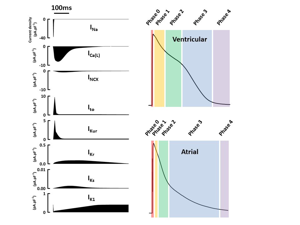
The journey starts with an action potential, the organized flow of charged ions like sodium (), potassium (), and calcium () across the cardiomyocyte membrane. Unlike the simple on-off switch of a nerve cell, the cardiac action potential has a distinct shape with a prolonged plateau phase. This plateau is critical. It's caused by the slow influx of calcium ions into the cell through specialized channels, and it's this very calcium that acts as the trigger for contraction.
Calcium's Chain Reaction
The small amount of calcium that enters the cell during the action potential isn't enough to cause a contraction on its own. Instead, it acts like a key to unlock a much larger reservoir. This is the principle of calcium-induced calcium release (CICR).
Inside each heart muscle cell is a specialized network of membranes called the sarcoplasmic reticulum (SR), which is packed with calcium ions. When the initial spark of calcium from the action potential arrives, it binds to and opens specific gates on the SR known as ryanodine receptors (RyRs). This causes a massive, rapid flood of calcium to pour out of the SR and into the cell's cytoplasm. It’s an amplification system, turning a small electrical signal into a powerful chemical one.
Think of it like this: the initial calcium influx is a single match, and the sarcoplasmic reticulum is a room full of fireworks. The match doesn't cause the explosion, but it ignites the fuse that does.
The Molecular Tug-of-War
With the cell now flooded with calcium, the stage is set for the mechanical action. The contractile machinery of the heart muscle is made of proteins called actin and myosin, arranged in repeating units called sarcomeres. In a resting state, their interaction is blocked by a regulatory protein duo: the s. Tropomyosin lies along the actin filament, covering the binding sites that myosin heads want to grab onto. Troponin holds the tropomyosin in place.
When calcium enters the scene, it binds to troponin. This binding causes a conformational change, a shift in shape, that pulls the tropomyosin strand aside. This exposes the myosin-binding sites on the actin filament. Now, the myosin heads can latch on, forming what's called a cross-bridge. Using energy from ATP, the myosin heads pull on the actin filaments, causing the sarcomere to shorten. This is the power stroke, the fundamental basis of muscle contraction. Millions of sarcomeres shortening in unison create the powerful squeeze of a heartbeat.
Excitation-contraction coupling refers to the sequence of events by which an AP (an electrical event) in the sarcolemma of the muscle cell initiates the sliding of the myofilaments, resulting in contraction (a mechanical event).
Connecting Signals to Squeezes
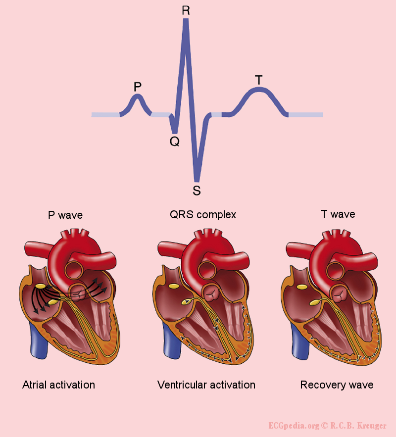
We can see this intricate dance play out on a surface electrocardiogram (ECG). The P-wave represents atrial depolarization, the electrical signal spreading through the heart's upper chambers. Shortly after, the atria contract (atrial systole), topping off the ventricles with blood.
Then comes the QRS complex, the large spike on the ECG. This signifies ventricular depolarization. It’s the electrical trigger for the main event. Following the QRS complex, the calcium flood begins, cross-bridges form, and the powerful ventricles contract (ventricular systole), pumping blood to the lungs and the rest of the body. The time interval between the electrical signal (QRS) and the resulting mechanical contraction is sometimes called the electromechanical window.
When this timing is off, problems arise. If the electrical activation is disorganized, as in some arrhythmias, the mechanical contraction becomes inefficient or chaotic. The ventricles may not have enough time to fill, or the contraction may be too weak to eject blood effectively. This highlights a fundamental truth of cardiology: electrical health is mechanical health. A breakdown in excitation-contraction coupling is a breakdown of the heart's essential function.
Time to check your understanding of this critical process.
What is the primary function of excitation-contraction coupling in heart muscle cells?
The initial, small influx of calcium () into a cardiomyocyte during an action potential is sufficient on its own to cause a full muscle contraction.
Understanding how a simple ionic signal translates into a powerful physical force is key to grasping both normal heart function and the mechanisms behind cardiac disease.