Advanced Biomechanics and Pathophysiology of the Hip Joint
Complex Joint Anatomy
The Acetabular Labrum
The hip joint's stability goes far beyond the fit of the femoral head into the acetabulum. A key player is the acetabular labrum, a ring of fibrocartilage that lines the rim of the socket. It's not just a passive bumper; it actively enhances joint stability.
The labrum deepens the acetabulum, but its most critical function is creating a suction seal. Think of it like a rubber gasket. It grips the femoral head, creating negative pressure within the joint. This suction is incredibly powerful, holding the ball firmly in the socket and resisting forces that would otherwise pull it apart.
This seal also ensures that synovial fluid, the joint's lubricant, is evenly distributed. By increasing the functional surface area of the acetabulum, the labrum helps spread the load of the body's weight across a wider area. This reduces peak pressure on the articular cartilage, protecting it from wear and tear.
The labrum deepens the socket and helps hold the joint together, providing crucial stability.
Deeper Stabilisers
Hidden within the joint capsule are two other important stabilising structures: the ligamentum teres and the zona orbicularis.
For a long time, the ligamentum teres was thought to be a vestigial remnant, an evolutionary leftover with no real function. We now know it's a vital intra-articular ligament. It acts as a strong but flexible tether connecting the femoral head directly to the acetabulum. This ligament is rich in nerve endings, giving it a proprioceptive role—it provides the brain with feedback on the hip's position and movement. While it carries a small artery that supplies some blood to the femoral head (more significant in children than adults), its primary role is mechanical stabilisation, especially in flexed and rotated positions.
The ligamentum teres acts as a proprioceptive check-rein, limiting extreme movements and providing sensory feedback.
Surrounding the narrowest part of the femoral neck is the zona orbicularis. This isn't a distinct ligament but rather a circular collar of deep fibres from the joint capsule. It functions like a buttonhole, constricting around the femoral neck and resisting distraction forces that could pull the head out of the socket. Its tight embrace significantly contributes to the hip's stability, working in concert with the labral seal.
The Capsular Ligaments
The hip joint is encased in a strong, fibrous capsule. This capsule is reinforced by three powerful ligaments that spiral around the joint: the iliofemoral, pubofemoral, and ischiofemoral ligaments.
Their spiral arrangement is biomechanically ingenious. When you stand up straight (hip extension), the ligaments twist taut, pulling the femoral head securely into the acetabulum. This provides immense passive stability, allowing you to stand with minimal muscular effort. Conversely, when you sit or bend (hip flexion), the ligaments unwind and become lax, permitting a wide range of motion.
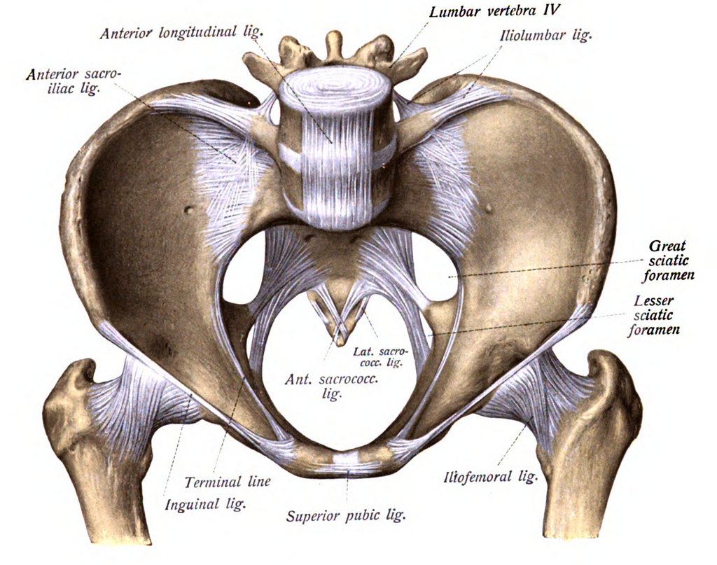
The iliofemoral ligament, also known as the Y-ligament of Bigelow, is the strongest ligament in the human body. It stretches from the ilium to the femur on the front of the joint and is the primary restraint against hyperextension.
The pubofemoral ligament is located anteroinferiorly and tightens during extension and abduction (moving the leg out to the side).
The ischiofemoral ligament, found on the posterior side, is the weakest of the three. It spirals up and over the joint, limiting internal rotation and extension.
Joint Health and Nutrition
The stability provided by these soft tissues is essential for the health of the joint itself. The inner surface of the joint capsule is lined by a synovial membrane, which produces synovial fluid. This viscous fluid serves two main purposes: lubrication and nutrition.
As a lubricant, it reduces friction between the articular cartilage of the femoral head and the acetabulum, allowing for smooth, painless movement. For nutrition, it's a lifeline. Articular cartilage has no blood supply, so it relies entirely on the synovial fluid for oxygen and nutrients. The loading and unloading of the joint during movement acts like a pump, circulating the fluid and forcing it into the cartilage.
When structures like the labrum or ligaments are damaged, the joint's mechanics are compromised. This instability can lead to abnormal movement patterns, disrupting the smooth circulation of synovial fluid and concentrating forces on small areas of cartilage. Over time, this can lead to cartilage breakdown and the development of osteoarthritis. Understanding these secondary stabilisers is therefore key to diagnosing and managing hip pathology.
Now, let's test your understanding of these complex hip structures.
What is the primary mechanism by which the acetabular labrum enhances hip joint stability?
How does the spiral arrangement of the three main capsular ligaments (iliofemoral, pubofemoral, ischiofemoral) contribute to stability?
The hip's intricate design balances incredible stability with a wide range of motion, thanks to these crucial soft tissue structures.