Echocardiography veiws
Welcome! I am glad you are here to learn about standard echocardiography views. Before we dive into the anatomy and the specific ultrasound windows, I would love to tailor our session to your needs.
What is your current background? Are you a medical student, a healthcare professional, or just curious about the topic?
I am a medical student.

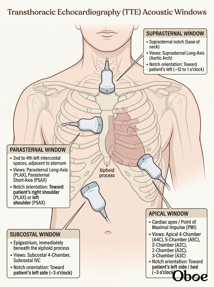
- Parasternal window: Located just to the left of the sternum (typically in the 2nd to 4th intercostal spaces).
- Apical window: Positioned over the cardiac apex (typically at the point of maximal impulse, or PMI).
- Subcostal window: Located just beneath the xiphoid process, using the liver as an acoustic window.
- Suprasternal window: Positioned in the dip of the neck just above the breastbone.
Show me a diagram of these 4 windows
I would be happy to show you how these four standard clinical windows map out on the patient's chest and abdomen.

Continue to the key structures visualized in the PLAX view
Let's focus on what you'll actually see on your screen during a (PLAX) scan. Because the ultrasound beam slices vertically from the front of the chest to the back, the structures closest to the probe appear at the top of your screen, while deeper structures sit at the bottom.
Anatomy of the PLAX Screen
- Right Ventricle (RV): Positioned at the very top of the sector because it is the most anterior chamber of the heart, lying directly behind the sternum.
- Interventricular Septum (IVS): The muscular wall separating the RV at the top from the left ventricle below it.
- Left Ventricle (LV): The main, large chamber sitting centrally. You will want to closely observe its thick muscular walls contracting toward the center.
- Mitral Valve (MV): Visible inside the LV, showing its anterior and posterior leaflets opening and closing like French doors.
- Left Atrium (LA): Located posteriorly and to the right side of the screen, acting as the reservoir that feeds into the LV.
- Left Ventricular Outflow Tract (LVOT) and Aortic Valve (AV): To the upper right of the LV, you will see the pathway leading out of the ventricle into the aortic root, where the delicate cusps of the aortic valve open and close.
- Descending Aorta: Seen as a small, circular cross-section at the very bottom of the screen, running posterior to the left atrium. This is a critical landmark to help you differentiate a pleural effusion from a .