Modern Radiology and Clinical Imaging Systems
Imaging Physics and Trade-offs
The Physics of Seeing Inside
Choosing the right medical imaging tool isn't guesswork. It's applied physics. Each modality—CT, MRI, and ultrasound—interacts with the body's tissues in a fundamentally different way. Understanding these interactions is the key to selecting the perfect tool for the diagnostic job, whether it's spotting a tiny fracture or a subtle change in soft tissue.
CT: A Numbers Game with Density
A Computed Tomography (CT) scan is essentially a sophisticated X-ray machine that rotates around the body, measuring how much radiation different tissues absorb. This property is called attenuation. Dense materials like bone block a lot of X-rays and have high attenuation, while air-filled lungs block very few.
To standardize this, CT data is converted into (HU), a quantitative scale that assigns a numerical value to the density of every single voxel, or 3D pixel, in the image. This turns a grayscale picture into a precise map of tissue densities.
| Substance | Hounsfield Units (HU) |
|---|---|
| Bone | +400 to +1000 |
| Soft Tissue | +40 to +80 |
| Blood (Fresh) | +30 to +45 |
| Water | 0 |
| Fat | -60 to -100 |
| Lung (Air) | -400 to -600 |
| Air | -1000 |
This numerical precision is CT's greatest strength. It excels at visualizing structures with large density differences, making it the go-to for identifying bone fractures, detecting acute bleeding in the brain (fresh blood is denser than brain tissue), and examining air-filled lungs. However, it struggles to differentiate between various soft tissues that have very similar HU values, like differentiating muscle from a tendon.
MRI: Listening to Protons
Magnetic Resonance Imaging (MRI) doesn't use ionizing radiation. Instead, it uses a powerful magnetic field to align the billions of hydrogen protons within your body's water molecules. A radiofrequency pulse then knocks these protons out of alignment. When the pulse stops, the protons relax back into alignment, releasing energy that the MRI scanner detects.
The magic of MRI lies in how these protons relax. Different tissues have different water content and molecular environments, causing their protons to relax at different speeds. The two key relaxation times are T1 and T2.
- T1 Relaxation (Longitudinal): This is the time it takes for protons to realign with the main magnetic field. In T1-weighted images, tissues where protons relax quickly appear bright. Fat is a prime example.
- T2 Relaxation (Transverse): This is the time it takes for the aligned protons to lose sync with each other after the pulse. In T2-weighted images, tissues where protons stay in sync for longer appear bright. Water and fluid are classic examples.
Think of it this way: T1 highlights fat. T2 highlights water (H2O).
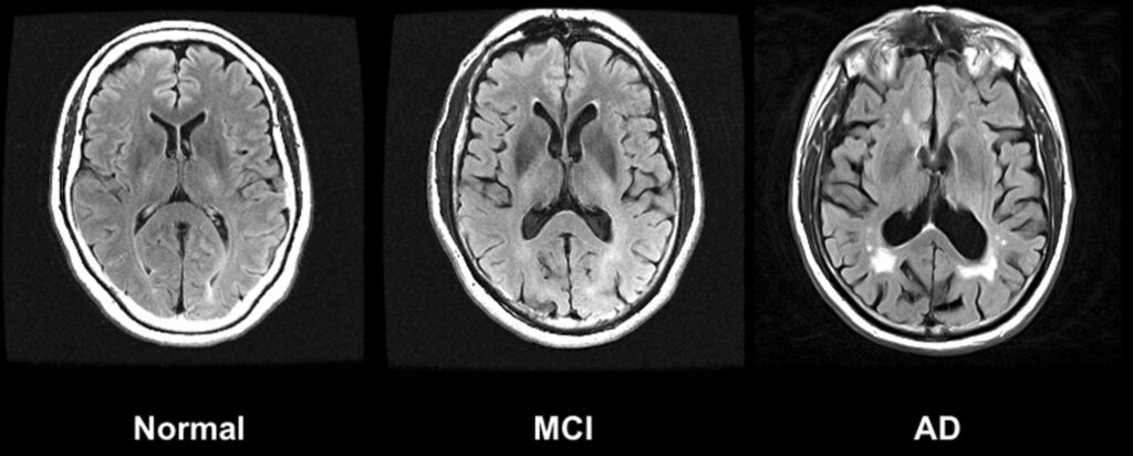
Radiologists can select different pulse sequences to emphasize these properties. A T1-weighted sequence is great for anatomy, as the contrast between fatty tissue and water-based structures is high. A T2-weighted sequence is ideal for pathology, as inflammation, tumors, and swelling (edema) all involve an increase in water content, making them shine brightly.
A common variation is the FLAIR sequence, which is like a T2 scan but with the signal from pure fluid (like cerebrospinal fluid) suppressed or nulled. This makes it much easier to see inflammation near fluid-filled spaces in the brain.
Ultrasound: The Echoes of Tissue
Ultrasound imaging is based on a completely different principle: sound waves. A transducer containing piezoelectric crystals converts electricity into high-frequency sound waves. These waves travel into the body and bounce off tissue boundaries, creating echoes. The same transducer then detects these returning echoes and converts them back into an electrical signal, which a computer uses to generate a real-time image.
The key physical property here is , which is a measure of how much a material resists the passage of sound waves. It's calculated as the product of the tissue's density () and the speed of sound within that tissue ().
The greater the difference in acoustic impedance between two adjacent tissues, the stronger the echo, and the brighter the boundary appears on the screen. This makes ultrasound fantastic for visualizing interfaces, like the boundaries of organs, blood vessels, and fluid collections. It is less effective where there are extreme impedance mismatches, such as with bone or air-filled lungs, which reflect nearly all the sound and create shadows.
Resolution and Noise Trade-offs
Every imaging modality faces a fundamental trade-off between spatial resolution and signal-to-noise ratio (SNR). Spatial resolution is the ability to distinguish two small, close objects as separate entities. It's determined by the size of the voxels (in CT/MRI) or the wavelength of the sound (in ultrasound). Smaller voxels mean higher resolution and sharper images.
However, smaller voxels also capture less signal (fewer photons for CT, fewer protons for MRI). This leads to a lower SNR, making the image appear grainy or noisy. To improve SNR, one might increase voxel size, but this sacrifices spatial resolution, potentially blurring fine details.
Radiologists constantly manage this balance. For a CT scan looking for a tiny lung nodule, high spatial resolution is critical, even if it means accepting more noise. For an MRI of the liver, where distinguishing subtle tissue changes is key, a higher SNR might be prioritized over razor-sharp edges.
Let's test your understanding of these physical principles.
A CT scanner produces images based on the varying degrees to which different tissues absorb X-ray beams. What is this physical property called?
A patient presents to the emergency room after a major car accident, and doctors suspect a bone fracture in the spine. Which imaging modality is the best choice for a quick and definitive diagnosis?
Mastering these core physics concepts provides the foundation for making informed clinical decisions. It's the difference between just looking at a picture and truly understanding the information it contains.