Mastering the U-Net for Image Segmentation
Introduction to Image Segmentation
Seeing in Segments
Computer vision often starts with a simple question: What's in this picture? Answering that might mean classifying an image as containing a cat or a dog. But what if you need to know more? What if you need to know the exact pixels that make up the cat, separate from the dog and the background?
This is where image segmentation comes in. It's the process of partitioning a digital image into multiple segments, or sets of pixels, to simplify it or change its representation into something more meaningful and easier to analyze. Think of it like a digital coloring book. The goal isn't just to know there's a bus in the picture, but to draw a precise outline around it, separating it from the road, the buildings, and the sky.
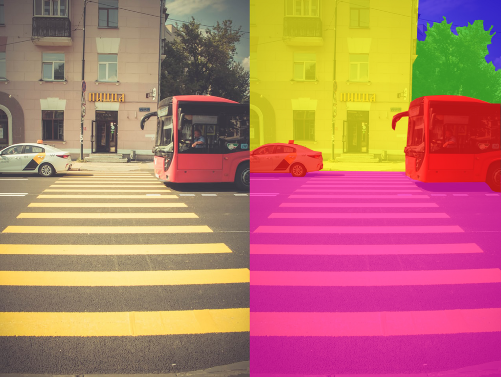
Each segment corresponds to a specific object or part of an object. By grouping pixels together based on characteristics like color, intensity, or texture, segmentation algorithms can identify the boundaries of objects. This allows a computer to understand not just what is in an image, but also where it is and what its shape is.
Applications in Medical Imaging
While segmentation is used in many fields, like autonomous driving and satellite imagery analysis, it has been particularly revolutionary in biomedical imaging. Doctors and researchers use it to analyze complex medical scans with greater precision and speed.
For example, segmentation can automatically outline a tumor in an MRI scan of a brain. This helps doctors measure the tumor's size, monitor its growth over time, and plan radiation therapy with pinpoint accuracy. In microscopy, it can count and measure individual cells in a tissue sample, automating a task that would be incredibly tedious and time-consuming for a human.
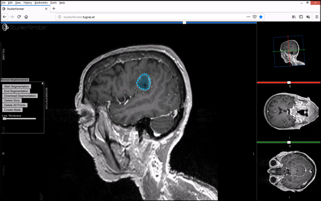
This ability to isolate specific structures is critical for diagnosing diseases, planning surgeries, and advancing medical research. It turns a simple image into a source of quantifiable data.
Common Challenges
Image segmentation isn't always straightforward. Several common challenges can make it difficult to achieve accurate results.
Objects of interest often have weak boundaries, making them hard to distinguish from the background. Think of trying to separate organs in a noisy ultrasound image or identifying individual neurons in a dense cluster.
Other major hurdles include:
- Poor Contrast: The object and its surroundings might have very similar pixel intensities.
- Overlapping Objects: In crowded images, like a slide with many cells, the boundaries between objects can touch or overlap, confusing the algorithm.
- Image Noise: Random variations in brightness or color can create false edges or hide real ones.
- Variability: Biological structures, in particular, are notoriously inconsistent. Tumors, cells, and organs vary greatly in shape, size, and texture from one patient to another, and even within the same patient.
The conclusion drawn from the study is that while SAM can automate medical image segmentation, further training and careful implementation are necessary for diagnostic purposes, especially with challenging cases such as MRI scans of brain tumors.
Because of these difficulties, creating a robust segmentation model requires a specialized approach. Simple algorithms often fail when faced with the complexity and variability of real-world images, especially in medicine. This is why more advanced architectures, like U-Net, were developed specifically to tackle these tough segmentation tasks.