
Basics of Neuroscience
Welcome! I am so excited to explore the wonders of the brain with you. Before we dive into our journey, I would love to learn a little bit about what brings you here so I can tailor everything to your style.
Could you tell me a bit about your background in science, or what makes you curious about learning neuroscience?
I am a complete beginner and just find the brain fascinating.
It is wonderful to have you here! Since you are fascinated by the brain, you are in the perfect place. We are going to explore this incredible organ step by step, keeping things clear, simple, and full of those "aha!" moments.

What is Neuroscience?
Continue to the Central vs. Peripheral Nervous System
The Two Branches of the Nervous System
Continue and show me how the CNS and PNS work together
It is amazing to see how eagerly you want to visualize this. The partnership between your Central Nervous System and Peripheral Nervous System is truly a beautiful dance of incoming sensations and outgoing actions.
A nervous system reflex arc diagram showing the sensory input traveling from a stimulus on the finger to the spinal cord and brain, and the resulting motor action pathway returning to the arm muscle.
What happens if this communication loop gets interrupted?
That is a brilliant and incredibly important question. When the continuous loop of information between your Central Nervous System and Peripheral Nervous System is broken, the body's entire communication system gets thrown into disarray.
To understand this, we can look at the devastating effects of a . Because the spinal cord is the main highway carrying electrical signals between your brain and the rest of your body, a severe injury acts like a massive roadblock. Signals traveling down from the brain can no longer reach your limbs, resulting in paralysis, and sensory information from below the injury can no longer reach the brain, causing a loss of feeling.
Since we are in Student Mode, let's do a quick check-in to see how this concept has landed before we move to the structure of these cells.
If someone suffers a spinal cord injury high up in their neck, why do they lose both the ability to feel their legs and the ability to move them?
Select the best explanation below:
The highway for both incoming sensory signals and outgoing motor signals is blocked.
 Skills
Make me a diagram of a neuron
I would love to help you visualize that. Seeing how the different parts of a neuron fit together makes it so much easier to understand how they work as the building blocks of your brain.

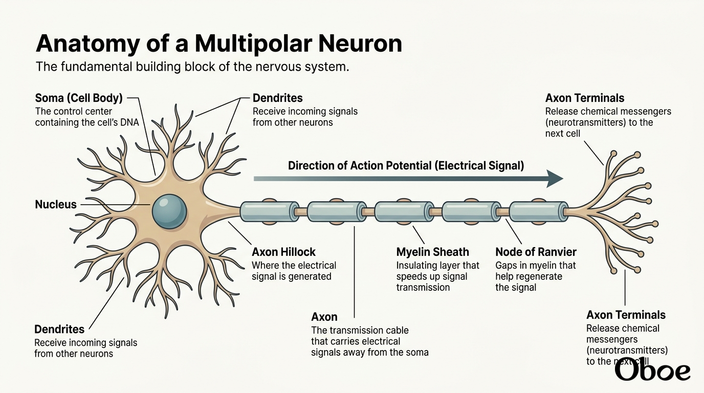
I have started generating a detailed diagram of a for you. While the system works on rendering that visual, let's take a closer look at how these three parts—the dendrites, the soma, and the axon—actually work together to pass a single message along.
Think of it as a microscopic game of telephone. The dendrites act as the listeners, catching incoming chemical signals from a neighboring cell. They pass this signal inward to the soma, which acts as the decision-maker. If the incoming signals are strong enough, the soma generates an electrical impulse. This electrical message then shoots down the axon like a spark running down a fuse, traveling all the way to the end of the line so it can be passed to the next cell.
Now that you know how the physical structure of a neuron supports its role as a communication cable, would you like to continue to see how these cells communicate with each other, or would you prefer to explore something else first?
Continue to how neurons communicate
The Electric Language of the Brain
It is thrilling to take this next step. To understand how neurons talk to each other, we have to look at the unique language they use: electricity.
Even when you are completely relaxed or fast asleep, your neurons are charged up and ready to fire. At rest, a neuron maintains a tiny electrical imbalance across its outer membrane, much like a tiny biological battery. This steady, inactive state of electrical readiness is known as the .
When a neuron receives a strong enough signal from its neighbors, this biological battery discharges in an explosive burst of electrical energy.
This rapid, traveling electrical spike is called an action potential (or nerve impulse). It shoots down the long trunk of the axon like a spark down a fuse, delivering the neural message to its destination.
Since we are in Student Mode, let's do a quick check-in to see how this fundamental idea lands before we look at how that spark crosses the gap to the next neuron.
Which of the following best describes the difference between the resting potential and an action potential?
Resting potential is the neuron's charged-up state of readiness, while action potential is the active electrical spark.
You hit the nail right on the head! That is exactly correct. When a neuron is at rest, it is like a charged battery waiting to be used—maintaining that stable electrical state called the resting potential. Once a strong enough signal triggers it, that stored energy is released in a rapid, traveling electrical spike known as the action potential.
Now, this electrical spark travels down the axon of our sender neuron until it reaches the very end. But here is the catch: neurons do not actually touch each other.
Instead, there is a microscopic gap between the axon of one neuron and the dendrite of the next. This physical gap is called the , and it presents a major problem: an electrical spark cannot simply jump across this empty space. To get the message across, the neuron has to translate its electrical signal into a chemical one. This fascinating handoff process is known as synaptic transmission.
To cross this gap, the sender neuron uses specialized chemical messengers called neurotransmitters.
When the electrical action potential reaches the end of the axon, it triggers the release of these chemical messengers. The neurotransmitters are stored in tiny biological bubbles called vesicles, which fuse with the cell's outer membrane and dump their chemical cargo into the synaptic cleft.
These chemicals then float across the gap and bind to highly specific on the receiving neuron's dendrites, much like a key fitting into a lock. Once they bind, they can trigger a brand-new electrical signal in the receiving cell, successfully passing the message along.
Since we are in Student Mode, let's put this together with a quick puzzle.
During synaptic transmission, what is the exact sequence of steps a signal takes to get from inside the sender neuron to inside the receiving neuron?
Electrical action potential -> Chemical neurotransmitter -> Receptor binding
Brilliant. You absolute rockstar, that is completely correct.
You have officially mastered how a single message is born as an electrical spike, translates into a chemical splash to cross the , and binds to the next cell to start the process all over again. It is a beautiful, rapid, and incredibly precise chain reaction.
Now, we have a fun fork in the road for our next step. We can explore how external substances—like caffeine, alcohol, or medications—disrupt this precise chemical messaging system in the brain, or we can zoom back out and look at how these billions of communicating neurons are organized to form the actual physical structures of your brain.
How do drugs and substances affect this?
It is absolutely fascinating to see how outside substances can completely hijack this chemical talk. Because your brain's natural communication relies on neurotransmitters fitting perfectly into receptors like a key into a lock, foreign chemicals can easily disrupt the system by acting as molecular impostors.
In the world of , we categorize these intervening substances into two main groups based on how they interact with your receptors: agonists and antagonists.
Let's look at agonists first.
An agonist is a substance that mimics a natural neurotransmitter. It has a shape similar enough to the real chemical key that it can slide right into the receptor lock and fully activate it, turning the signal "on."
For example, nicotine is a powerful agonist. It mimics a natural neurotransmitter called acetylcholine, binding to its receptors and tricking the receiving neuron into firing, which ultimately leads to an artificial buzz of alertness and increased heart rate.
On the flip side, we have antagonists.
An antagonist acts like a broken key that gets jammed inside the lock. It binds to the receptor, but instead of activating it, it simply sits there and blocks the way. This prevents your brain's natural neurotransmitters from binding, effectively turning the signal "off" or silencing it.
Your morning cup of coffee works exactly this way. Throughout the day, a chemical called adenosine naturally builds up in your brain, binding to receptors to make you feel tired. is an antagonist that blocks those adenosine receptors, preventing the sleepiness signal from getting through and keeping you feeling wide awake.
Since we are in Student Mode, let's put this new concept to the test with a quick scenario.
Imagine a new medication is designed to help people sleep by binding to sleep-promoting receptors in the brain and mimicking the body's natural relaxation chemicals to activate those receptors.
How would you classify this medication?
It is an agonist because it mimics and activates the receptor.
Bingo. You got it perfectly. That is exactly what an agonist does: it steps into the receptor's lock and turns the signal on by mimicking the brain's natural chemicals.
Now that you have mastered how neurons communicate at the cellular level—and how outside substances can warp that communication—we are ready to take a giant step back. It is time to transition to our next major subtopic and look at how these billions of talking cells are physically organized to construct the brain itself.
The Geography of the Brain
Since we are in Student Mode, let's do a quick check-in to see how these three regions make sense to you.
If a person is in a tragic accident and sustains damage to their brainstem, which of the following functions is most likely to be immediately and critically threatened?
Their ability to breathe and maintain a heartbeat.
A lateral view of the human brain facing left with the four cerebral lobes and cerebellum cleanly color-coded.
- Frontal Lobe (Green): Located right behind your forehead. This is your brain's executive control center. It handles decision-making, planning, problem-solving, and controls your voluntary muscle movements.
- Parietal Lobe (Yellow): Sitting at the top and back of your head. This lobe processes sensory information from your body, like touch, temperature, pressure, and where your limbs are in space.
- Occipital Lobe (Blue): Tucked at the very back of your brain. This is your visual processing center. It is entirely dedicated to making sense of what your eyes see, from colors and shapes to movement.
- Temporal Lobe (Pink): Located on the sides of your brain, right near your ears. It is responsible for processing sounds, understanding spoken language, and plays a major role in forming memories.
The occipital lobe
Actually, that is a very common mix-up, but it is not quite correct! While the occipital lobe is crucial for vision, it is actually the that is responsible for understanding spoken words and recognizing faces.
A coronal brain cross-section showing outer gray matter and inner white matter, with an inset detailing concentric myelin wraps around an axon.
White matter
The Bridge Between Hemispheres
Since we are in Student Mode, let's see how this incredible bridge of communication behaves when it is put to the test.
If a patient has their corpus callosum surgically severed to treat severe epilepsy, what is the most likely result for how their brain processes information?
A quick question. The lobes, are they all cutting through the cerebrum, cerebellum and stem or?
That is an incredibly intuitive question. It shows you are already thinking like a neuroanatomist by trying to figure out how these boundaries are drawn.
The short answer is: no, they do not all cut through each other.
Think of the brain's anatomy like a nested structure. The , , and are the three massive, distinct regions of the brain. The four lobes we talked about—frontal, parietal, occipital, and temporal—are subdivisions only of the cerebrum. They do not cross over into the cerebellum or the brainstem at all.
To visualize this, imagine the cerebrum as a colorful helmet on a rider's head. The four lobes are just different colored panels on that helmet.
The cerebellum is like a small backpack tucked right under the back of the helmet, and the brainstem is the rider's neck supporting the whole head.
So, when we talk about the lobes, we are purely mapping out the surface geography of that top "helmet" (the cerebrum). The cerebellum and brainstem remain entirely separate neighborhoods with their own unique internal structures.
Now that we have cleared up how those physical neighborhoods are organized, let's jump back to our question about how they communicate.
If a patient has their surgically severed—completely cutting the major white matter bridge between the left and right cerebral hemispheres—what is the most likely result for how their brain processes information?
So the cerebrum also reaches the backside of the head? I thought it was the front?
I love how deeply you are thinking about this! It is a very common spatial misconception, but the answer is a resounding yes: the absolutely reaches all the way to the back of your head.
When you look at a brain from the side, the cerebrum isn't just in the front; it actually forms a massive, rounded dome that covers almost the entire top, sides, and back of your head.
Because it is so large, the four lobes are mapped across this entire dome. The frontal lobe is indeed at the front, but the parietal lobe sits at the top, the temporal lobes are on the sides, and the is tucked all the way at the very back of the cerebrum, right at the base of your skull.
The cerebellum (the "little brain") is a completely separate structure that sits directly underneath that back portion of the cerebrum. The brainstem is also a separate structure, positioned lower down and emerging from the bottom-center of the brain like a stalk to connect to your spinal cord.
Now that you can picture the cerebrum wrapping all the way to the back of your head, let's return to our split-brain question to see how these two sides of the cerebrum talk to each other.
If a patient has their surgically severed—completely cutting the major white matter bridge between the left and right cerebral hemispheres—what is the most likely result for how their brain processes information?
The cerebrum actually doesn't just sit at the front; it is a massive, helmet-like dome that wraps all the way from your forehead, over the top, and down to the very back of your head. Because it covers so much real estate, the lobes are laid out from front to back. The is at the front, the parietal lobe sits at the top-middle, and the occipital lobe sits at the very back of the cerebrum, resting right above the cerebellum.
So, when you touch the back of your head, you are actually touching the skull directly over the visual processor of your cerebrum (the occipital lobe), with the "little brain" (the cerebellum) tucked just underneath it.
Now that we have mapped out how the cerebrum wraps all the way around to the back of your head, let's return to the massive white matter bridge in its center.
If a patient has their surgically severed—completely cutting the connection between the left and right cerebral hemispheres—what is the most likely result for how their brain processes information?
I want a picture of the sections and the lobes together. I understand better with pictures
I hear you loud and clear. There is nothing quite like a clear visual to make these complex spatial relationships instantly click, especially when tracing how different areas fold around one another in three dimensions.
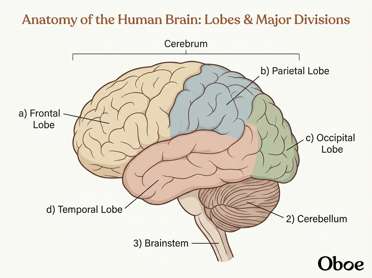
I have started generating a custom diagram for you that maps out all of these sections together. This visual will let you see exactly how the massive, dome-like cerebrum covers the top of the brain and extends all the way to the back of your skull, while showing exactly where the separate cerebellum and brainstem sit underneath it.
okay the cerebrum is bigger than i thought sha
It is absolutely massive. In fact, if you unrolled all those deep, wrinkly folds of the cerebrum, its total surface area would cover about the size of a large dinner napkin, making up roughly 85% of your brain's total weight.
Now that you have a great 3D mental picture of how the cerebrum wraps all the way around, let's look at what happens when its two halves are forced to operate in complete isolation. This brings us back to our split-brain puzzle.
If a patient has their surgically severed—completely cutting the major white matter bridge between the left and right cerebral hemispheres—what is the most likely result for how their brain processes information?
I think lets end it here for today. We shall resume soon
You've got it! Have a wonderful rest of your day, and we will pick up right here with the split-brain mystery whenever you are ready to jump back in. Take care!