Clinical Anatomy of the Trochanteric Anastomosis
Arterial Network Overview
The Hip's Vascular Crossroads
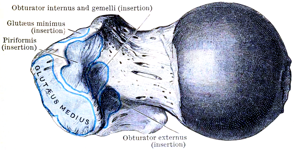
Tucked away in a small depression on the femur called the trochanteric fossa is a critical meeting point for several major arteries. This network, known as the trochanteric anastomosis, forms a vital hub for blood supply to the hip joint. An anastomosis is like a traffic circle for your circulatory system, where multiple arterial roads connect, providing alternative routes for blood flow. This redundancy is crucial, especially for supplying the head of the femur.
This network ensures the femoral head receives a steady supply of oxygenated blood, even if one of the contributing vessels is compromised. The main supply line to the femoral head comes from the retinacular arteries, which are delicate branches that arise primarily from this anastomosis.
The Four Key Players
Four primary arteries converge to form the trochanteric anastomosis. Their paths, especially in relation to the piriformis muscle, are key anatomical landmarks.
- Superior Gluteal Artery (SGA): This artery enters the gluteal region by passing above the piriformis muscle. It then sends a branch down to join the network.
- Inferior Gluteal Artery (IGA): In contrast, the IGA travels below the piriformis muscle to contribute to the anastomosis.
- Medial Circumflex Femoral Artery (MCFA): Often considered the most important contributor, the MCFA wraps around the femur from back to front, providing the main source for the retinacular arteries that directly feed the femoral head.
- Lateral Circumflex Femoral Artery (LCFA): This artery travels along the outer side of the hip and sends an ascending branch up to connect with the other vessels.
Remembering the relationship with the piriformis is simple: the Superior Gluteal Artery goes superior (above), and the Inferior Gluteal Artery goes inferior (below).
Clinical Significance
The trochanteric anastomosis is more than just an interesting anatomical feature; it's a critical backup system. In the event of a blockage in one of the main arteries, this collateral circulation can sometimes maintain blood flow to the femoral head.
However, this network has its limits. In a displaced femoral neck fracture, the retinacular arteries, which are the final, direct supply to the head, are often torn. The collateral flow from the anastomosis may not be enough to prevent avascular necrosis, a condition where the bone tissue dies due to a lack of blood supply. Understanding this vascular map is therefore essential for orthopedic surgeons when treating hip fractures and other injuries.
This highlights the outsized role of the circumflex arteries, especially the MCFA, in nourishing the most vulnerable part of the hip joint.
What is the primary function of the trochanteric anastomosis?
Which artery travels above the piriformis muscle to join the trochanteric anastomosis?
By understanding the convergence of these four arteries and their collective role, you can better appreciate the intricate design and potential vulnerabilities of the hip's blood supply.