Clinical Anatomy of Intercostal Veins
Posterior Venous Layout
The Posterior Venous Network
The posterior intercostal veins are responsible for draining the back of the thoracic wall. While their arterial counterparts have a relatively straightforward pattern, the venous side has a few unique twists, especially at the highest levels.
The 1st posterior intercostal vein on each side is an outlier. Instead of joining the larger systems below, it charts its own course. On both the right and left, it ascends over the neck of the first rib to drain directly into the corresponding or sometimes the vertebral vein.
Things get more coordinated just below that. The 2nd, 3rd, and sometimes the 4th posterior intercostal veins join together to form a single trunk on each side: the superior intercostal vein.
However, their final destinations differ. The right superior intercostal vein has a short, direct path, draining into the arch of the as it curves over the root of the right lung. The left superior intercostal vein takes a longer route, traveling across the aortic arch to empty into the left brachiocephalic vein.
Position in the Groove
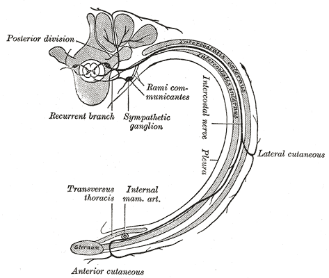
The arrangement of vessels and nerves within the costal groove of each rib follows a consistent, superior-to-inferior order. This pattern is crucial for clinical procedures like thoracentesis (draining fluid from the chest) or placing a chest tube.
The order from top to bottom is Vein, Artery, Nerve. A helpful mnemonic is VAN.
The posterior intercostal vein is the most superior structure in the bundle, tucked safely into the deepest part of the costal groove. The intercostal artery lies just below it, and the intercostal nerve is the most inferior and most exposed of the three.
This predictable anatomy is why needles and tubes are always inserted over the superior border of a rib. This placement avoids the neurovascular bundle located along the inferior border, minimizing the risk of hemorrhage or nerve damage.
The Remaining Veins
From the 4th intercostal space down to the 11th, the drainage pattern becomes more consistent, though still asymmetric.
On the right side: The posterior intercostal veins from spaces 4 through 11 drain directly into the azygos vein as it ascends through the posterior mediastinum.
On the left side: The drainage is split. Veins from spaces 4 through 8 typically drain into the accessory hemiazygos vein. Veins from spaces 9 through 11 drain into the hemiazygos vein. Both of these veins then cross the midline to empty into the larger azygos vein.
Understanding this venous map is essential for interpreting imaging studies of the chest and for planning surgical approaches to the posterior thoracic wall. The asymmetry and variability are key features of this system.
Which structure is located most superiorly within the costal groove of a typical rib?
To safely perform a procedure like placing a chest tube, the instrument should be inserted just over the superior border of a rib.