Aortic Dissection Explained
Aortic Anatomy
The Body's Main Artery
The aorta is the largest artery in your body, the main highway for blood leaving the heart. Think of it as the trunk of a massive tree, originating from the left ventricle—the heart's primary pumping chamber. From there, it extends down to your abdomen, branching out along the way to deliver oxygen-rich blood to every part of your body.
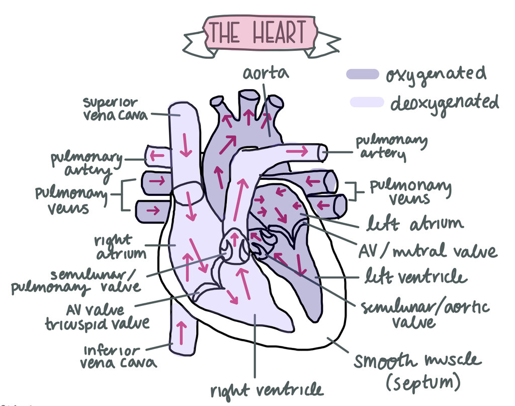
This powerful vessel is responsible for systemic circulation. When the left ventricle contracts, it pushes blood into the aorta with tremendous force. The aorta's elastic walls expand to accommodate this surge and then recoil, which helps maintain blood pressure and ensures a steady flow of blood to the brain, organs, and limbs.
A Journey in Segments
The aorta isn't just one long, straight tube. It's a continuous vessel with four distinct segments, each with a specific location and function.
-
Ascending Aorta: This is the initial section, rising out of the heart's left ventricle. The coronary arteries, which supply blood directly to the heart muscle, branch off right at its start.
-
Aortic Arch: As its name suggests, this segment arches over the heart, like the handle of a cane. Three major arteries branch off from the arch to supply blood to your head, neck, and arms.
-
Descending Thoracic Aorta: After the arch, the aorta travels down through your chest (the thorax). Along the way, it sends smaller branches to the ribs, chest muscles, and organs like the esophagus and lungs.
-
Abdominal Aorta: Once it passes through the diaphragm (the large muscle separating the chest from the abdomen), it becomes the abdominal aorta. This segment is crucial for supplying blood to the major abdominal organs, including the stomach, liver, intestines, and kidneys. It finally ends by splitting into two iliac arteries, which continue down into the legs.
The Three-Layered Wall
The aortic wall is not just a simple tube; it's a complex, three-layered structure designed to withstand high pressure and remain flexible. Each layer has a unique role.
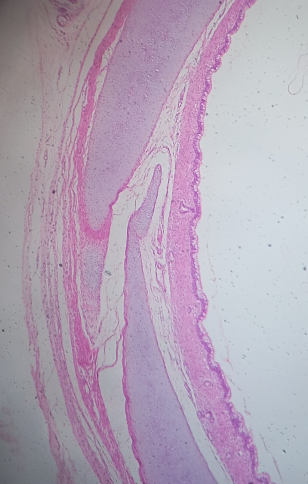
From the inside out, the layers are the tunica intima, tunica media, and tunica adventitia.
Tunica Intima: This is the innermost layer, a thin, smooth lining of endothelial cells that comes into direct contact with blood. Its slick surface minimizes friction, allowing blood to flow easily and preventing clots from forming.
Tunica Media: The middle layer is the thickest and most important for the aorta's function. It's packed with smooth muscle and a high concentration of elastic fibers. These elastic fibers allow the aorta to stretch with each heartbeat and then snap back, helping to propel blood forward. The smooth muscle allows the vessel to contract or relax, regulating blood pressure and flow.
Tunica Adventitia: This is the tough, fibrous outer layer. It's made of collagen and other connective tissues that provide structural support and prevent the aorta from over-expanding. It also contains tiny blood vessels (the vasa vasorum) that supply the aortic wall itself with oxygen and nutrients, as well as nerves that help regulate its function.
Let's check your understanding of the aorta's structure and function.
Which chamber of the heart does the aorta originate from?
What is the primary function of the elastic fibers within the tunica media, the aorta's middle layer?
Understanding the aorta's anatomy—its segments, layered wall, and central role in circulation—is key to appreciating how vital this vessel is for overall health.