
Basics of Neuroscience
Welcome! I'm so excited to explore the wonders of the human brain and nervous system with you.
To help me tailor our journey to your exact needs, tell me a little bit about what brings you to neuroscience today. Are you studying for an upcoming course, pursuing a personal curiosity, or looking to apply it to a specific field like psychology, medicine, or artificial intelligence?
I want to apply it to my professional field
To tailor this neuroscience journey perfectly to your goals, I'd love to know what kind of professional field you are in so we can connect these neural systems directly to your day-to-day work.
Biotechnology and Bioinformatics
It's wonderful to have you here! Because you work in biotechnology and bioinformatics, we'll make sure to bridge these foundational biological concepts directly to how data is generated, modeled, and targeted in your industry.

The Master Division of the Nervous System
| System Division | Key Components | Primary Biotech/Bioinformatics Relevance |
|---|---|---|
| Central Nervous System (CNS) | Brain and Spinal Cord | High-throughput target discovery for neurodegenerative diseases |
| Peripheral Nervous System (PNS) | Cranial, spinal, and peripheral nerves | Neuromuscular drug modeling, bioelectronic medicine, and wearables |
In bioinformatics, this distinction is crucial because the biological data profiles generated by these two systems are wildly different. For example, high-throughput single-cell RNA sequencing data will reveal entirely different gene expression patterns and receptor profiles in CNS tissue compared to a peripheral nerve biopsy.
Before we dive into the specific roles of the brain and spinal cord, do you have any initial thoughts on how this high-level division might affect the computational models or data types you work with?
Let's continue to the key functions of the brain, spinal cord, and peripheral nerves.
Let's zoom in on how these divisions actually share the workload to keep you running. Every second, your nervous system coordinates an elegant, three-way relay between the brain, the spinal cord, and your peripheral nerves.
The Neural Information Relay
To understand how they work together, imagine stepping on a sharp Lego brick. This triggers a lightning-fast chain of events:
- The Peripheral Nerves (The Sensors): Specialized sensory receptors in your foot detect the mechanical pressure and instantly generate an electrical signal. This signal travels up your leg via the peripheral nerves.
- The Spinal Cord (The Local Router & Fast Responder): Before the signal even reaches your brain, it enters your spinal cord. For immediate dangers, the spinal cord can execute a completely on its own, sending an automatic motor command back down to pull your foot away.
- The Brain (The Chief Executive): Meanwhile, the spinal cord also routes the signal up to the brain. Your brain processes the data, calculates the emotional response (annoyance), stores the memory of where you stepped, and decides what to do next (like cleaning up the floor).
A three-step reflex arc showing sensory input from a foot, local processing in a spinal cord cross-section, and ascending signaling toward the brain.
I've started creating a visualization of this reflex pathway to help you see exactly how the signal splits between the spinal cord's local loop and the pathway climbing up to the brain.
From a data perspective, the spinal cord is much more than a simple conduit; it's a critical preprocessing hub. It filters, prioritizes, and compresses sensory streams before they reach the brain's main processing centers.
Next, we'll look at the major structural regions of the brain itself—the cerebrum, cerebellum, and brainstem—where this upstream data is ultimately delivered and analyzed.
Continue to the major brain regions: cerebrum, cerebellum, and brainstem
Let's dive straight into the brain itself, which is divided into three major structural regions: the cerebrum, the cerebellum, and the brainstem.
The Brain's Core Geography
- The : This is the massive, wrinkled outer dome of the brain. It is the central processing unit for high-level cognitive functions, decision-making, sensory integration, and voluntary movement.
- The : Tucked neatly under the back of the cerebrum, this structure translates complex motor plans into smooth, coordinated physical actions.
- The Brainstem: This is the structural trunk connecting the brain to the spinal cord, responsible for hardcoding and automating basic survival functions like breathing, heart rate, and sleep cycles.
To bring this into a biotech and bioinformatics context, think of these regions as possessing unique cellular profiles and genetic programs. If you were analyzing a single-cell RNA-sequencing dataset from the brainstem, you would find high expression of genes related to automatic homeostasis, whereas a cortical sample from the cerebrum would show highly complex networks of genes dedicated to synaptic plasticity.
I have initiated a diagram to help visualize where these three key regions are structurally situated relative to each other.
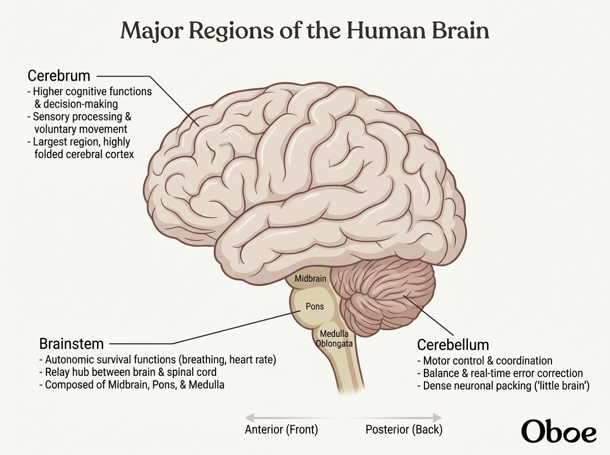
With this structural roadmap of the nervous system complete, we've wrapped up our first major milestone! We're ready to zoom in to the micro-level next and explore the individual cells that make this computing network possible: neurons and glial cells.
Continue to Neurons, Glia, and Cellular Structure
Welcome to the cellular world of the nervous system! Now that we have mapped out the macro-geography of the brain and spinal cord, we are ready to zoom all the way down to the micro-level to meet the specialized processors that make this complex computational engine run: the .
Anatomy of a Neuron
- The Soma (The CPU): This is the cell body of the neuron. It contains the nucleus and the cell's metabolic machinery. In our computer analogy, this is where the processing cores live, synthesizing the proteins and energy needed to keep the cell alive and deciding whether to pass on an incoming signal.
- The Dendrites (The Input Ports): Branching out like tree limbs from the soma, these structures act as receivers. They catch incoming chemical messages from neighboring cells and channel them inward to the cell body.
- The Axon (The Transmission Cable): Once the soma decides to fire, it sends a single electrical impulse down this long, slender cable. Axons can stretch over massive distances, sometimes running all the way from your spinal cord down to your big toe.
- The Axon Terminal (The Output Port): Located at the very end of the axon, this structure converts the electrical signal back into a chemical message, releasing neurotransmitters across the synaptic gap to communicate with the next cell's input ports.
- The (The Insulating Wrap): This is a protective, fatty coating wrapped tightly around the axon. Just like the plastic insulation on a USB cable, myelin prevents electrical signals from leaking out, allowing data to zip down the axon up to a hundred times faster than it would on a bare, uninsulated nerve fiber.
A classic multipolar neuron visual showing dendrites, soma, myelin sheath, and axon terminals.
I have created an interactive visualization of this basic anatomy to help you see exactly how a single neuron is structured to capture, process, and propagate information down its line.
From a bioinformatics and modeling perspective, neuron morphology is incredibly diverse. Different brain regions utilize entirely different shapes of neurons (such as pyramidal cells in the cerebrum vs. purkinje cells in the cerebellum) to handle their unique processing workloads. Translating these complex 3D shapes into digital morphological skeletons is a major area of work in modern neuroanatomy pipelines.
Before we look at the 'other half' of the brain's cellular population—the glial support cells—let's test our understanding of how signal processing moves through a single neuron.
Soma -> Dendrites -> Axon -> Axon Terminal
 Skills
You hit the nail on the head! While you correctly identified the structures, the actual signal flow goes in the other direction: it is received by the Dendrites (the input ports), passes through the Soma (the processor), zips down the Axon (the transmission cable), and exits via the Axon Terminal (the output port).
Now, let's turn our attention to the other half of the brain's cellular workforce. For a long time, neurons got all the glory in neuroscience, while glial cells (or glia) were dismissed as mere biological glue holding the processors in place. Today, we know they make up about half of the cells in your brain and perform active computational, protective, and maintenance roles that are highly relevant to biotechnology and clinical therapeutics.
Meet the Glial Support Network
In our computer system analogy, if neurons are the silicon chips, glial cells are the cooling fans, power supplies, security software, and maintenance technicians that keep the server rack from crashing. Let's look at the four major types of glial cells:
- : Shaped like stars, these cells are the ultimate utility players. They physically wrap around brain blood vessels to help form the highly selective blood-brain barrier. They maintain the chemical environment around neurons, clean up excess neurotransmitters, and even regulate local blood flow to match neural activity.
- : These are the myelin makers of the Central Nervous System. A single oligodendrocyte can stretch out its lipid-rich arms to wrap myelin sheaths around the axons of up to 50 nearby neurons, acting as a centralized insulation factory.
- : These perform the exact same myelination job as oligodendrocytes, but exclusively in the Peripheral Nervous System. Unlike their CNS cousins, a single Schwann cell wraps itself entirely around just one segment of a peripheral axon.
- : These are the resident immune cells of the brain. Since the blood-brain barrier blocks standard immune cells from entering, microglia act as specialized, mobile scavengers, patrolling the tissue to engulf cellular debris, damaged cells, and foreign pathogens.
In the biotechnology industry, glial cells have transitioned from background players to prime therapeutic targets. For example, understanding how microglia transition from protective scavengers to chronic drivers of neuroinflammation is a massive area of focus for drug discovery in neurodegenerative diseases like Alzheimer's and ALS.
Before we look at how researchers computationally model these complex, branched cellular structures, let's see how well you can distinguish between these crucial support cells. If you were analyzing a therapeutic drug designed specifically to repair damaged myelin in a peripheral nerve in the leg, which glial cell would you target?